Photobiomodulation/Low Level Laser Therapy

Keyhole

Ambassador
Ambassador
FOTCM Member
This thread's been a long time coming. There have been several discussions about light therapy, so I figured it would be best to have a thread focused on it. From what I understand, the terms photobiomodulation and low level laser therapy can be used interchangeably. They essentially both refer to the use of specific wavelengths of light as a clinical tool to help heal and reverse disease. Light has effects both on the surface of the skin and can penetrate deeply into the tissues. It stimulates mitochondrial energy production, cell protection and detoxification.

In my opinion the most interesting effects of light seem to be its interaction with biological water, whereby it induces water structuring which: possibly provides activation energy for chemical reactions to take place, is responsible for the flow of lymph, blood and other fluid, and may provide a physical/electrical barrier to prevent unwanted solutes and ions from entering the cell.

Nonetheless, here is a good introductory article on the topic of red light therapy:

The Therapeutic Effects of Red and Near-Infrared Light (2015)

"Penetrating red light is possibly the fundamental anti-stress factor for all organisms. The chronic deficiency of such light is, I think, the best explanation for the deterioration which occurs with aging."
- Raymond Peat

1. Preface

About two years ago, I spent a lot time reading Ray Peat's articles, trying to make sense of his various ideas regarding health. In many of his articles, Peat wrote that red light is healthy and even crucial for well-being, because it can activate mitochondrial respiration.

For example, in his article Aging Eyes, Infant Eyes, and Excitable Tissues (2006), he wrote:
"Old observations such as Warburg’s, that visible light can restore the activity of the 'respiratory pigments,' showed without doubt that visible light is biochemically active. By the 1960s, several studies had been published showing the inhibition of respiratory enzymes by blue light, and their activation by red light."

Peat didn't give many references to justify his claims, but after doing some searches on PubMed, I realized that there are literally thousands of papers supporting his views.

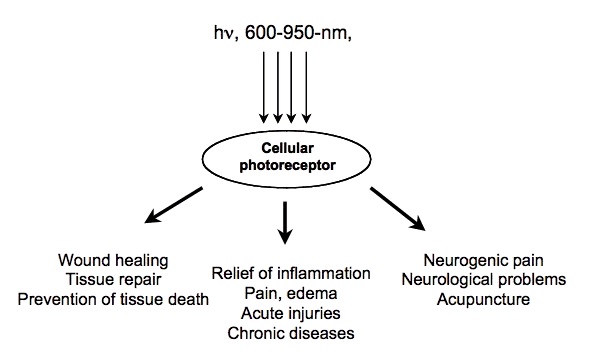
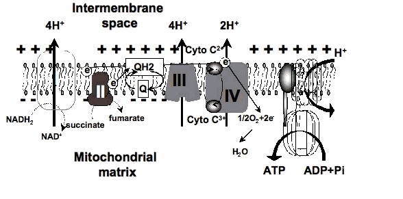
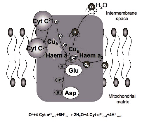
2. How could red light improve metabolism?

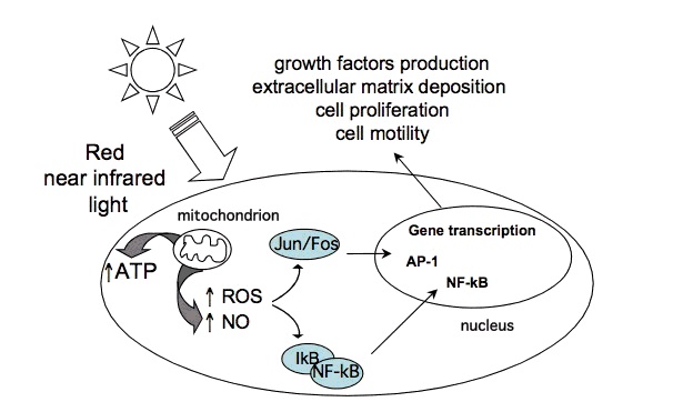
Certain wavelengths of electromagnetic radiation directly increase energy (ATP) in the tissues, and the activation of cytochrome c oxidase (Cox), the mitochondrial respiratory enzyme discovered by Nobel laureate Otto Warburg, seems to be the main mechanism.[1-6] On the molecular level, red light seemingly causes the photodissociation of nitric oxide (NO). When the cells are stressed, cells can produce NO which can bind to Cox, inhibiting its ability to bind oxygen.[2,7-11]

The following citations are from a news article on Nature (2006):

"Recent findings suggest that the enzyme [Warburg] identified, cytochrome oxidase, is a key player in a new understanding of how the cell's energy metabolism affects health and disease. And surprisingly they show that light has a profound effect on how the enzyme works — and could even be used to treat degenerative disease." [...]
"According to cell biologist Salvador Moncada of University College London, evolution really has crafted cytochrome oxidase to bind not only oxygen but also NO. 'One effect of slowing respiration in some locations is to divert oxygen elsewhere in cells and tissues,' he says. This prevents oxygen levels sinking dangerously low." [...]
"We have shown that light can indeed reverse the inhibition caused by NO binding to cytochrome oxidase, both in isolated mitochondria and in whole cells," says biochemist Guy Brown, at the University of Cambridge, UK. "And what's more, we found that light can protect cells against NO-induced cell death." [11]


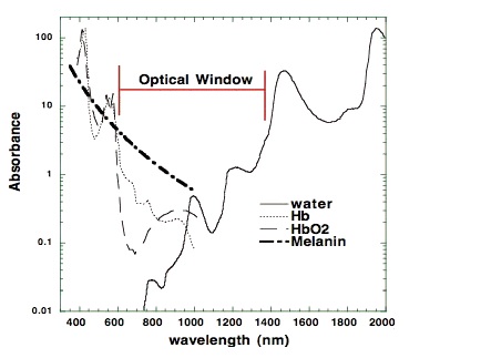
The most relevant wavelengths seem to be 600-1070nm --- in other words, red light and the penetrating shorter wavelengths of near-infrared radiation (NIR).[1,2]Our eyes are able to see 400-700nm radiation (blue, green and red light). Therapeutic effects come from 600-1070nm radiation (red and near-infrared light). Within this range, some wavelengths seem to have better effects than others. For example, in one study, 665nm and 810nm rays were beneficial, while 730nm and 980nm were not.[12]

There are important differences in the penetrating power of the different wavelengths. Visible red light doesn't penetrate tissue very well, but near-infrared does that quite well. If near-infrared is directed to the skin, the power seems to decrease 1000-fold by every 2-3 centimetres. However, a worm study shows that even those very small doses seem to have an effect on the cellular level.[13-15] Later in this article, I will also show that light apparently also has an systemic effect, which probably spreads through the circulation to the whole body. When it comes to Peat's claims about blue light, high intensities can inhibit the same enzyme (Cox), and this can lead to retinal damage and other problems.[11,16]

3. The health effects of red light and near-infrared radiation: The research history

John H. Kellogg's book (1910) is freely available on Archive.org website.

John Kellogg and incandescent lamp therapy (1910): The therapeutic use of red light is not a new phenomenon. The very earliest reports on the topic have been published in the 19th century, the most well-known article being The Red Light Treatment of Small-Pox (1895) by Niels Finsen, who also got the 1903 Nobel Prize in medicine, for his research regarding the health effects of light (especially ultraviolet radiation).[17]

In 1910, John Harvey Kellogg published his 200-page book Light Therapeutics, which included a large amount of information about the therapeutic usefulness of light therapy with incandescent light bulbs and arc lights. According to his book, light therapy can be effectively used for diabetes, obesity, chronic fatigue, insomnia, baldness, cachexia and many other health problems.[18] Similar therapeutic usage was also reported in Margaret A. Cleaves' book Light energy, its physics, physiological action and therapeutic applications (1904) and Leopold Freund's book Elements of general radio-therapy for practitioners (1904).

In the next decades after Kellogg's, Cleaves' and Freund's books, the information was apparently forgotten. However, some elderly people have told me that in their youth, some doctors used to recommend infrared lights for some therapeutic purposes (such as pain after dental operations).

The invention of laser light (1960-): In the beginning of 60's, the first red-light emitting lasers were invented. A couple of years after that, Endre Mester from Hungary reported that red laser light could improve hair growth on mice. Quickly after that it was also found that red laser light could accelerate wound healing in animals - and in '70s the first human trials were being done.[2] (At the same time, Marta Fenyo started focusing on polarized light therapy after working with Mester.)

In the '80s and '90s most of the research was conducted in the Soviet Union. There was many kinds of effects studied. For example, some studies reported protective effect against x-rays and other studies focused on heart disease.[19-22] Modern study of light/LLLT: In the Western countries, research on red light didn't really start before the 21th century, and most of the interesting studies have been released within the last five years (2010-2015). A dramatic amount of clinical research is being conducted nowadays: In 2014, more than 400 PubMed titles on the topic were published.

In most of the studies on red light or near-infrared therapy, a low-power laser device (output often less than 100mW) has been used, therefore the widely used abbreviation LLLT (low-level laser therapy).Many studies have also been conducted with LED's or other light sources. One can find also relevant studies by using keywords such as narrow-band light therapy, phototherapy, photobiomodulation, photobiostimulation, low-level light therapy, laser acupuncture, water-filtered infrared-A, near-infrared light, visible light, polarized-light therapy, or light-emitting diode irradiation. Despite different ways of generating the light, the wavelength used in the studies always fits between 600 and 1070 nanometres, because that is the effective range.

The studies have most often been conducted using a monochromatic light source (eg. 630, 633, 660, 670, 780, 808, 810, 890, 904, 940, 1064 or 1072nm). The parameters such as light dose, power output, pulsing or diameter of the light ray usually varies in the different studies because different devices are being used. There is no general consensus on which parameter is the most reliable, but many have yielded positive results.

4. The health effects of red light and near-infrared radiation: The extent of research

Today, PubMed keyword LLLT returns more than 4000 scientific papers. Hundreds of clinical trials have been conducted, and dozens of systematic reviews can also be found.

Here is a list of some health issues that have been reported to improve by light therapy (usually low-level laser). In some cases, the effects are still disputed, while other results are confirmed by meta-analyses.

Photobiomodulation indications (human studies):
Achilles tendinitis
Muscle pain
Acne
Neuropathic foot ulcer
Age-related macular degeneration
Neck pain
Aphthous ulcer
Nipple pain
Bell’s palsy
Oral mucositis
Body contouring
Orthodontic pain
Bone fractures (hand)
Osteoarthritis
Burn scars
Postherpetic neuralgia
Carpal tunnel syndrome
Pressure ulcer
Chronic joint disorders
Radiation dermatitis
Dentin hypersensitivity
Raynaud’s phenomenon
Depression
Restenosis
Erythema
Rheumatoid arthritis
Fibromyalgia
Shoulder tendinopathy
Frozen shoulder
Skin ageing
Hair loss
Sternotomy (recovery)
Hand-foot-and-mouth disease
Stroke
Herpes labialis
Temporomandibular disorders
Hypothyroidism
Tennis elbow
Low back pain
Venous leg ulcer
Lymphedema
Wound healing
Muscle growth
Xerostomia


Many animal studies have also been conducted:
Achilles tendinitis
Laryngitis
Acne
Liver cirrhosis
Acute pain
Liver regeneration
Adipose tissue inflammation
Lung injury
Age-related macular degeneration
Muscle injury
Allergic asthma
Muscle loss (sarcopenia)
Alzheimer’s disease
Nerve injury
Arthritis
Osteomyelitis
Atherosclerosis
Methanol toxicity (eyes)
Auditory neuropathy
Multiple sclerosis
Bone graft incorporation
Myocardial infarct
Bone fracture
Myonecrosis
Burns
Neuropathic pain
Cancer
Opiate addiction
Colitis
Oral mucositis
COPD
Osteoarthritis
Dentin regeneration
Osteoporosis
Depression
Periodontitis
Diabetes (eyes)
Parkinson’s disease
Diabetes (kidney)
Pressure ulcer
Endophthalmitis
Radiation damage
Exercise performance
Rheumatoid arthritis
Hair loss
Spinal cord injury
Hearing loss
Stroke
Heart failure
Temporomandibular inflammation
Hemarthrosis
Thrombocytopenia
Hyperalgesia
Tinnitus
Hypertension
Traumatic brain injury
Kidney fibrosis
Wound infection
Kidney injury

5. The health effects of red light and near-infrared radiation: A few examples of the clinical study results

5.1. Age-related macular degeneration


German clinicians conducted a retrospective analysis of 200 elderly subjects with age-related macular degeneration (most of them also had cataracts). The subjects were treated using a LLLT device emitting near-infrared light (780nm). The light was targeted to their eyes, through the conjunctiva.[65] The subjects were treated four times during two weeks. Placebo group was given a mock treatment. In the LLLT group, the visual acuity was improved in 95% of the subjects. Most of them were able to see a few rows lower on the Snellen chart. The improved vision was maintained for 3-36 months after treatment. LLLT also appeared to improve edema, bleeding, metamorphosia, scotoma and dyschromatopsia in some patients.

5.2. Knee Osteoarthritis

Hungarian researchers studied the use of near-infrared LLLT in knee osteoarthritis patients, in a double-blinded placebo controlled trial (830nm).[73] Intervention group got infrared treatment on their affected joint twice a week, over a period of four weeks. The placebo group got a similar treatment of 100-fold lower intensity.

In the intervention group, the pain scores were (on a scale from 1 to 10):
- 5.75 before the treatment
- 1.71 after the last treatment session
- 1.18 two months after completing the therapy


In the placebo group, the pain scores were:
- 5.62 before treatment
- 4.13 after the last treatment session
- 4.12 two months after completing the therapy

Some other studies on this issue haven't been nearly as successful, but as discussed below, it might be related to dose parameters or some other methodological factors in the studies.

5.3. Labial herpes

The researchers of University of Vienna Medical School studied the usage of red light on labial herpes in a double-blind, placebo-controlled trial.[61] There was a 12-fold difference in the median time until the recurrence of herpes symptoms between the groups.(Schindl&Neumann 1999) The subjects were treated in a recurrence-free period. The intervention group were treated for 10 minutes daily for two weeks with visible red light (low-level laser). Placebo group got a similar treatment, but the laser wasn't turned on. The subjects wore masks, so that they couldn't see whether they were given the real treatment. The patients were instructed to return to the department at the time of symptom recurrence. The median recurrence-free interval in the laser-treated group was 37.5 wk compared with 3 wk in the placebo group.

5.4. Wisdom teeth extraction

A 2013 Italian study focused on patients who had their lower third molar removed surgically. The patients were assigned to a LLLT or control group. The active group received near-infrared light to the extraction site and on cheek (980nm).[92] The pain level and edema were measured 24h after the surgery. The subjects in the active group had much less pain and edema compared to the control group. The patients receiving low-level laser therapy reported that their level of pain was 3.75/10 on the next day after surgery. In the control group, the score was 7.1/10.
A couple of other studies on this issue have been published. If we look at the all of the data critically, it isn't completely conclusive, but still very promising.

5.5. Hypothyroidism

This 2013 randomized, placebo-controlled study involving 43 patients with Hashimoto's thyroiditis-induced hypothyroidism was conducted in São Paolo, Brazil.[37] The active group received ten treatment sessions (twice a week) involving the near-infrared irradiation of the skin area close to their thyroid glands (830nm). The placebo group received red light treatment of very low intensity[37] After 10 treatment sessions, the thyroxine (T4) medication was discontinued in both groups. During the next 9 months, the medication was slowly re-introduced if the thyroid hormone levels didn't normalize without medication.

In the control group, hypothyroidism remained after the discontinuation of T4 and the final dosage after the reintroduction of medication was actually higher than before the discontinuation.

However, in the active group, 48% of the patients maintained normal thyroid hormone levels without any medication. The rest also could decrease their dosage a little.

Average T4 dose in the active group (baseline -> 9 months): 93µg -> 39µg
Average T4 dose in the placebo group (baseline -> 9 months): 90µg -> 107µg

In the active group, some other positive changes were also noticed (thyroid volume, TPO antibodies and echogenicity index).

In my personal opinion, this is a very remarkable result. The Brazilian researcher had also conducted a pilot study before this randomized study, with equally positive results. Similar effects have also been reported in some Russian and Ukrainian dissertations (not translated to English).[36-43]

If you are interested in this topic, see also my more recent article Hypothyroidism: Could it be treated with LIGHT?.

6. The systemic anti-inflammatory effect

Usually the red/near-infrared is applied locally to the treatable tissue. If the patient suffers from knee osteoarthritis, then the light is going to be shone on the knee, and so on.

However, light also has systemic effects which seem to be transmitted mainly by circulation of blood. The researcher Natalya Zhevago has conducted an interesting study, in which the patients got some visible light and infrared to the sacral area (low back).[86] The light was quite similar to sunlight, except that it didn't contain UV radiation or blue light, and the infrared portion was polarized. According to one study, polarization of light enchances the metabolic effect slightly.[87] The subjects' blood was analyzed after the treatment. The results were interesting. Subjects' pro-inflammatory cytokines (TNF-α, IL-6 etc.) were dramatically reduced in the subjects, especially in those with initially high values. Also, the concentrations anti-inflammatory cytokines increased.[86] A dramatic decrease in the level of pro-inflammatory cytokines TNF-α, IL-6, and IFN-γ was revealed: at 0.5 h after exposure of volunteers (with the initial parameters exceeding the norm), the cytokine contents fell, on average, 34, 12, and 1.5 times[...]

Another research group reported that polarized light protects rabbits from atherosclerosis when targeted on the outside surface of ear.[88]
"Lovastatin (0.002% in diet) or daily 5-min or 20-min PLT [polarized-light therapy] on the outside surface of ears was started 2 weeks after induction of hypercholesterolemia. [...] [T]he anti-atherosclerotic activity of PLT was superior to lovastatin: 5-min and 20-min PLT decreased the plaque area to 42.2% and 26.4%, respectively." "Importantly, 5-min PLT also exerted a remarkable efficacy in LDL reduction"

The possible mechanisms of these systemic effects hasn't been studied widely yet. One research group conducted an interesting in vitro study and suggested that the effect might be mediated by some growth factors.[89] In the present study, increased growth factors by indirect irradiation stimulate the ERK phosphorylation in the presence of LPS. Especially, indirect 635 nm irradiation can affect MAPK activation and is correlated with the inhibition of pro-inflammatory cytokine expression. The effects observed by Zhevago were quite opposite to the typical effects of UV radiation, which increases TNF-α, IL-6 and other pro-inflammatory cytokines. Sun-exposure is also related to increased IL-6 levels.[90,91]

In human studies, large doses of IL-6 and TNF-α have been demonstrated to suppress peripheral thyroid hormone metabolism by decreasing T3 and increasing rT3.[92,93] We could also speculate, whether lack of sufficient therapeutic light could be one cause of the "rT3 dominance" and hypothyroid symptoms. In two studies, half of the hypothyroid patients getting near-infrared treatment did not require any medication through the 9-month follow-up after the treatment period, somewhat establishing the importance of light for thyroid health.[36,37] Moreover, in a Russian study (Kovalyova 2002), the diabetic patients' total cholesterol was apparently reduced from 7.98 to 5.31 in one month, a change also seen in thyroid treatments.[32-35,94]

7. Light sources (laser, LED, light bulbs, heat lamps, sunlight, therapeutic devices)

"Many people who came to cloudy Eugene to study, and who often lived in cheap basement apartments, would develop chronic health problems within a few months. Women who had been healthy when they arrived would often develop premenstrual syndrome or arthritis or colitis during their first winter in Eugene." - Ray Peat

Laser and LED: Nowadays red light and near-infrared are studied mostly with low-level laser devices (LLLT), but many researchers also use light-emitting diodes (LED) or other light sources. The coherence and pulsing of light (Laser vs. other sources): Although most of the researchers have been using coherent light (laser), it seems that coherence is not a requirement for the beneficial effects. This is logical considering the studies utilizing light-emitting diodes. The idea is also supported by Kellogg's reports, which were based on the usage of incandescent bulbs and arc lights.[2,95] However, it is possible that non-coherent light sources might work best with different parameters than laser (power density, total energy, etc.). According to some data I've seen, laser might reach deep tissues better than other types of light. Laser devices also often emit pulsed light, but it's not clear whether pulsing is very meaningful.[96]

Here are some quotes from an accomplished Estonian LLLT researcher Tiina Karu:

"[T]he stimulative action of various bands of visible light at the level of organisms and cells was known long before the advent of the laser. Also, specially designed experiments at the cellular level have provided evidence that coherent and noncoherent light with the same wavelength, intensity, and irradiation time provide the same biological effect [11-13]. Successful use of LED's in many areas of clinical practice also confirms this conclusion."-Tiina Karu (2003)

"According to action spectra, optimal wavelengths are 820-830, 760, 680, and 620 nm. Large volumes and deeper layers of tissues can successfully irradiated by laser only (e.g. inner and middle ear diseases, injured siatic or optical nerves, deep inflammations etc.). The LED's are excellent for irradiation of surface injuries." -Tiina Karu (year?)

Sunlight: When I was writing my Circadian Rhythms essay, I used to think about the possible explanations of the therapeutic effects of walking outdoors. Sunlight can increase the production of vitamin D and it can also suppress melatonin, but now we have a brand new mechanism that could explain why it's good to spend time outdoors.

There is an interesting correlation between latitude and mean blood cholesterol levels.(Grimes et al. 1996)

A review article on this subject states that in central Europe, the amount of IR-A radiation is limited to 20mW/cm2, which is actually quite a good amount compared to the power output of usual laser devices. However, the sunlight is not monochromatic, which probably increases the dose requirements.[1] Especially in Northern countries, it seems that sunlight is correlated to better health. In some countries, the average cholesterol level of population depends largely on the season. In the Great-Britain, for example, the changing of winter into summer has been shown to decrease cholesterol by 0.8mmol/L (30mg/dl). Also, there seems to be a significant correlation between latitude and cholesterol levels.[97-102]
"In all groups, we found a strong seasonal effect, with 5%–10% higher [total cholesterol] concentrations in winter compared to the summer."[102]

While providing a significant amount of terapeutic wavelengths, sunlight also contains some harmful UV-radiation and blue light, which might decrease the benefits a little bit. Incandescent, halogen and heat lamps: These types of indoor lamps also provide a significant amount of red light and near-infrared. Some of these lamps have internal reflectors, so the light is targeted at one direction. Heat lamps by Philips or Osram have quite a good spectrum with low amount of blue light, but a large amount of their power is emitted as warming IR-B radiation, and only ~12% of the power is emitted as the therapeutic wavelengths (600-1070nm). However, the heat lamps are often high power (up to 250W), so they still emit quite a significant amount of therapeutic wavelengths and might have health effects.

4100K halogen lamp seems to be good source of the therapeutic wavelengths. Sunlight and incandescent/heat lamps also provide significant amounts of 600-1070nm light. (Image source: Heelspurs)

Incandescent lamps and especially halogen lights seem to emit quite a significant amount of their energy as therapeutic wavelengths (up to 35%). They are also quite cheap, which might make them a wise choice for light therapy. In my country, the price of Osram halogen lamps (30-40W) with reflector is about 5€ (~5-6 dollars).

However, there is probably no scientific research testing these kinds of lamps, so it's not proven whether they really work. The preliminary reports by Kellogg were very promising, but as I already mentioned, nowadays almost every research group uses either laser or LED, probably because they don't emit any heat and they are monochromatic or at least the bandwidth is quite narrow.

Energy saving lamps: Because of the phase-out of incandescent lamps, it will soon be increasingly difficult to buy incandescent lamps. It's somewhat sad that they are going to be replaced with compact fluorescent lamps (CFL), which emit some UV but only low amounts of protective red and near-infrared light. This is the reason why some of the researchers, such as Richard Funk and Alexander Wunsch, who also appeared in the Bulb Fiction documentary, have stated that increase in the CFL usage might be harmful to eyes.

Infrared saunas: The possible benefits of infrared saunas aren't usually based on this aforementioned mechanism, because even the shortest-wave infrared saunas don't go below 1400nm. However, the warming effect of far-infrared might also have some beneficial effects.[103]

Infrared lamps:
In theory, a cheap incandescent or halogen lamp with an internal reflector could provide all the pro-metabolic effects, yet nobody has studied the issue (since Kellogg). Some lamps are being sold as "infrared lamps". For example, Beurer models IL30 and IL50 seem to be quite popular models in my country. According to one source, they emit 780-1400nm wavelengths, so they might be suitable. However, no scientific data exists on these.

Therapy devices: The good aspect of lasers and LED lights is that the wavelength can be narrowly chosen (monochromatic light). For example, 830nm has proven good in many studies, and the wavelength is invisible to the eyes. Therefore it'd theoretically make a good and non-disturbing therapy device.

Here is a list of some commercial light devices, some of which have been used in LLLT/light therapy studies: Anodyne, Bioptron, HairMax LaserComb, Omnilux, Noveon NaiLaser, Biolight, Quantum Warp, Syrolight BioBeam, HIRO 3.0, Picasso Lite, HELBO® TheraLite Laser, Super Lizer, BioPhotas Celluma, TinniTool EarLaser and Mustang 2000.

The problem with commercial devices is the fact that they are expensive yet a typical halogen spot lamp (eg. Osram Classic Superstar R50 or R63) could theoretically be as good therapeutically, since the lamps also provide a great amount of the healthy wavelengths.

8. Does it really work? Trying to understand the contradictory results:


Negative results: Even though most of the studies on LLLT have reported positive effects, there have been many studies in which LLLT didn't bring any better results than placebo.

Swedish researchers Jan Tuner and Lars Hode have published an article It's All in the Parameters: A Critical Analysis of Some Well-Known Negative Studies on Low-Level Laser Therapy, in which they explained why, in many cases, the negative results can be explained by the bad parameters used in the study.[104-106]

One typical factor is using a very low dose or power density (eg. 0.004J, 0.005J/cm2), though in some cases too high dose might also block the beneficial effect. Sometimes the problem might arise from targeting the light at the wrong place, using non-effective wavelenghts (eg. 730, 980nm), using the patient as his own control (not taking the systemic effect into account), using an inappropriate control group, or using a malfunctioning device (see below).

One year ago, I tried to discuss some of the LLLT studies with one scientist who works with the doctors who write national recommendations on medical treatments of various diseases. We looked on studies, in which they treated osteoarthritis with LLLT. Most of the results were positive, but in one study condicted in University of Ottawa didn't report a beneficial effect even though the parameters seemed generally "good" on the paper. I couldn't explain this effect to the officer, but later I understood that the light dose they gave to the patients was quite low (0.12J per joint). This is a possible explanation for the negative results of this study.[105,107]

Osmangazi University also conducted a study with negative results.[108] The parameters were similar to the aforementioned Hungarian study with great results.[73] I couldn't find a simple explanation for the negative result, but it could be related to the fact that the diameter of the laser ray was 1mm, and if such a small ray is directed at the wrong point of the joint, then there might be no effect. In one systematic review it was noted that usually the studies using greater ray had better results.[73,108,109]

"Lack of evidence": LLLT isn't still a widely known subject, and therefore many probably think there is no research on the subject. However, a PubMed search with keyword LLLT provides 4000 results, and many meta-analyses have already been published.

Here are some examples of the recent systematic reviews and/or meta-analyses:

Breast cancer-related lymphedema (2015)
Exercise performance and recovery (2015)
Oral mucositis (2014)
Pain relief (2010)
Shoulder tendinopathy (2015)
Temporomandibular disorders (2015)


However, despite the existence of many high-quality RCT trials and good meta-analyses, there is also a really huge amount of problematic research with a high risk of methodological bias. Because of some low-quality studies, some people think that all research on LLLT is bad:

"Like acupuncture, there is a huge literature (4000 on the Pubmeds) of mostly poorly done studies, some showing effect, some not. The Cochrane reviews were not supportive of laser therapy, but note the studies are uniformly lousy."

I think the quality problem is most obvious in the older studies, while nowadays the research has better quality standards. In an 2014 meta-analysis examining the effects LLLT on oral mucositis, most of the studies published after 2011 were of high quality, while older studies had more methodological issues.[72]

"Like acupuncture, better studies demonstrate decreasing effects."

This is also untrue. In a recent meta-analysis on LLLT and shoulder tendinopathy, the average PEDro score of in the studies reporting positive results was 7.5, while the score was 7.6 in the studies reporting negative results.[152]

Also, half of the studies reporting negative results were conducted using a malfunctioning laser device (Roland Pagani) that emitted less than 1% of the output power stated by the manufacturer. Therefore, it can be actually said the studies that reported positive effects were actually better quality.

When I tried to read Wikipedia article on LLLT, I found it difficult to understand the claim that LLLT "may be mildly effective, but in most cases no better than placebo in relieving [...] osteoarthritis, [...] acute and chronic neck pain", since the systematic reviews referenced in Wikipedia article were showing moderate-quality evidence of the beneficial effect.[110-112]

Meta-analyses: When reading meta-analyses on LLLT, some problems might arise. One problem is that so much research is being published nowadays, that there usually exists new data on the subject of the meta-analysis you're reading.

Also, the studies are very heterogenic because the different research groups are using different parameters (wavelength, power, power density, pulsing, dose, treatment time). In one systematic review, in one study patients received 0.3J of light energy per joint, while in another study the dose was 480J per joint (1600-fold difference). Only the latter study reported positive results. However, this doesn't mean that higher energy is better in every case. Some studies have reported a biphasic dose-response curve.[7,113]

In some meta-analyses, there might also be problems related to cherry picking or data synthesis.[114]

9. Light therapy: Animal studies

Red light and near-infrared have also been extensively studied using different animal models. Here is a short list of some research on the subject. The results have been generally very positive:

Rats:
The 2011 review article is mostly focused on how light could affect brains. Most of the research data is from animal studies. (Rojas&Gonzalez-Lima 2011)
Acute joint inflammation [115]
Bone metabolism [116]
Burns [117-120]
Cortical metabolic capacity and memory retention[121]
Diabetic retinopathy [122]
Diabetic kidney [123]
Heart failure-related inflammation [124]
Hypertension [125]
Kidney injury [126]
Laryngitis [127]
Lung injury [153]
Methanol-induced eye injury [128]
Myocardial infarction (infarct size) [129]
Palatal wound healing [130]
Peripheral nerve regeneration [131]
Reflux laryngitis [132]
Rheumatoid arthritis [133]
Skeletal muscle injury [134]
Tendon healing [135]
Traumatic brain injury [136]
Zymosan-induced arthritis [137]


Mice:
Encephalomyelitis [138]
Snake venom poisoning [139]
Traumatic brain injury [140]


Dogs:
Hair regrowth (non-inflammatory alopecia) [141]
Myocardial infarction [142]
Recovery from surgery (hemilaminectomy) [143]
Sperm motility [144]


Rabbits:
Embolic stroke [145]
Wound healing [146]


10. Conclusions

Nowadays, the knowledge of the physiological effects of light is mainly limited to blue light's effects on circadian rhythm, yet the importance of red and near-infrared light is also likely to be a very important factor. Research indicates that red light and near-infrared appear to have quite a wide range of therapeutic effects. Light is very cheap to produce, so there is a possibility that in future, red light would be used as a very cost-effective treatment for various wounds, injuries and chronic diseases.


Link to the article here
 
Heres an older article published in 2013 on the mechanisms behind photobiomodulation. (Note: a LOT of research has been published in the past four years, so the human/animal article is not be 100% up to date)


MECHANISMS OF LOW LEVEL LIGHT THERAPY -Michael R. Hamblin

The use of low levels of visible or near-infrared (NIR) light for reducing pain, inflammation and edema, promoting healing of wounds, deeper tissues and nerves, and preventing tissue damage has been known for almost forty years since the invention of lasers. Originally thought to be a peculiar property of laser light (soft or cold lasers), the subject has now broadened to include photobiomodulation and photobiostimulation using non-coherent light. Despite many reports of positive findings from experiments conducted in vitro, in animal models and in randomized controlled clinical trials, LLLT remains controversial. This likely is due to two main reasons; firstly, the biochemical mechanisms underlying the positive effects are incompletely understood, and secondly, the complexity of rationally choosing amongst a large number of illumination parameters such as wavelength, fluence, power density, pulse structure and treatment timing has led to the publication of a number of negative studies as well as many positive ones. In particular, a biphasic dose response has been frequently observed where low levels of light have a much better effect than higher levels.

This introductory review will cover some of the proposed cellular chromophores responsible for the effect of visible light on mammalian cells, including cytochrome c oxidase (with absorption peaks in the NIR), and photoactive porphyrins. Mitochondria are thought to be a likely site for the initial effects of light, leading to increased ATP production, modulation of reactive oxygen species, and induction of transcription factors. These effects in turn lead to increased cell proliferation and migration (particularly by fibroblasts), modulation in levels of cytokines, growth factors and inflammatory mediators, and increased tissue oxygenation. The results of these biochemical and cellular changes in animals and patients include such benefits as increased healing of chronic wounds, improvements in sports injuries and carpal tunnel syndrome, pain reduction in arthritis and neuropathies, and amelioration of damage after heart attacks, stroke, nerve injury, and retinal toxicity.

1. HISTORY

In 1967, a few years after the first working laser was invented, Endre Mester in Semmelweis University, Budapest, Hungary wanted to test if laser radiation might cause cancer in mice [1]. He shaved the dorsal hair, divided them into two groups and gave a laser treatment with a low powered ruby laser (694 nm) to one group. They did not get cancer, and to his surprise the hair on the treated group grew back more quickly than the untreated group. This was the first demonstration of "laser biostimulation". Since then, medical treatment with coherent-light sources (lasers) or noncoherent light (light-emitting diodes, LEDs) has passed through its childhood and adolescence. Currently, low-level laser (or light) therapy (LLLT), also known as "cold laser", "soft laser", "biostimulation" or "photobiomodulation" is practiced as part of physical therapy in many parts of the world. In fact, light therapy is one of the oldest therapeutic methods used by humans (historically as solar therapy by Egyptians, later as UV therapy for which Nils Finsen won the Nobel prize in 1904 [2]). The use of lasers and LEDs as light sources was the next step in the technological development of light therapy, which is now applied to many thousands of people worldwide each day. In LLLT, the question is no longer whether light has biological effects, but rather how energy from therapeutic lasers and LEDs work at the cellular and organism levels, and what are the optimal light parameters for different uses of these light sources.

One important point that has been demonstrated by multiple studies in cell culture [3], animal models [4] and in clinical studies is the concept of a biphasic dose response when the outcome is compared with the total delivered light energy density (fluence). It has been found that there exists an optimal dose of light for any particular application, and doses lower than this optimum value, or more significantly, larger than the optimum value will have a diminished therapeutic outcome, or for high doses of light a negative outcome may even result. Evidence suggests that both energy density and power density are key biological parameters for the effectiveness of laser therapy, and they may both operate with thresholds (i.e., a lower and an upper threshold for both parameters between which laser therapy is effective, and outside of which laser therapy is too weak to have any effect or so intense that the tissue is inhibited) [5].

The reason why the technique is termed LOW-level is that the optimum levels of energy density delivered are low when compared to other forms of laser therapy as practiced for ablation, cutting, and thermally coagulating tissue. In general, the power densities used for LLLT are lower than those needed to produce heating of tissue, i.e., less than 100 mW/cm2, depending on wavelength and tissue type.

2. PHYSICAL MECHANISMS

According to quantum mechanical theory, light energy is composed of photons or discrete packets of electromagnetic energy. The energy of an individual photon depends only on the wavelength. Therefore, the energy of a "dose" of light depends only on the number of photons and on their wavelength or color (blue photons have more energy than green photons, that have more energy than red, that have more energy than NIR, etc). Photons that are delivered into living tissue can either be absorbed or scattered. Scattered photons will eventually be absorbed or will escape from the tissue in the form of diffuse reflection. The photons that are absorbed interact with an organic molecule or chromophore located within the tissue. Because these photons have wavelengths in the red or NIR regions of the spectrum, the chromophores that absorb these photons tend to have delocalized electrons in molecular orbitals that can be excited from the ground state to the first excited state by the quantum of energy delivered by the photon. According to the first law of thermodynamics, the energy delivered to the tissue must be conserved, and three possible pathways exist to account for what happens to the delivered light energy when low level laser therapy is delivered into tissue.

The commonest pathway that occurs when light is absorbed by living tissue is called internal conversion. This happens when the first excited singlet state of the chromophore undergoes a transition from a higher to a lower electronic state. It is sometimes called "radiationless de-excitation", because no photons are emitted. It differs from intersystem crossing in that, while both are radiationless methods of de-excitation, the molecular spin state for internal conversion remains the same, whereas it changes for intersystem crossing. The energy of the electronically excited state is given off to vibrational modes of the molecule, in other words, the excitation energy is transformed into heat.

The second pathway that can occur is fluorescence. Fluorescence is a luminescence or re-emission of light, in which the molecular absorption of a photon triggers the emission of another photon with a longer wavelength. The energy difference between the absorbed and emitted photons ends up as molecular vibrations or heat. The wavelengths involved depend on the absorbance curve and Stokes shift of the particular fluorophore.

The third pathway that can occur after the absorption of light by a tissue chromophore, represents a number of processes broadly grouped under an umbrella category of photochemistry. Because of the energy of the photons involved, covalent bonds cannot be broken. However, the energy is sufficient for the first excited singlet state to be formed, and this can undergo intersystem crossing to the long-lived triplet state of the chromophore. The long life of this species allows reactions to occur, such as energy transfer to ground state molecular oxygen (a triplet) to form the reactive species, singlet oxygen. Alternatively the chromophore triplet state may undergo electron transfer (probably reduction) to form the radical anion that can then transfer an electron to oxygen to form superoxide. Electron transfer reactions are highly important in the mitochondrial respiratory chain, where the principal chromophores involved in laser therapy are thought to be situated. A third photochemistry pathway that can occur after the absorption of a red or NIR photon is the dissociation of a non-covalently bound ligand from a binding site on a metal containing cofactor in an enzyme. The most likely candidate for this pathway is the binding of nitric oxide to the iron-containing and copper-containing redox centers in unit IV of the mitochondrial respiratory chain, known as cytochrome c oxidase (see below).

It should be mentioned that there is another mechanism that has been proposed to account for low level laser effects on tissue. This explanation relies on the phenomenon of laser speckle, which is peculiar to laser light. The speckle effect is a result of the interference of many waves, having different phases, which add together to give a resultant wave whose amplitude, and therefore intensity, varies randomly. Each point on illuminated tissue acts as a source of secondary spherical waves. The light at any point in the scattered light field is made up of waves that have been scattered from each point on the illuminated surface.If the surface is rough enough to create path-length differences exceeding one wavelength, giving rise to phase changes greater than 2Pi.jpg, the amplitude (and hence the intensity) of the resultant light varies randomly. It is proposed that the variation in intensity between speckle spots that are about 1 micron apart can give rise to small but steep temperature gradients within subcellular organelles such as mitochondria without causing photochemistry. These temperature gradients are proposed to cause some unspecified changes in mitochondrial metabolism

3. BIOCHEMICAL MECHANISMS

There are perhaps three main areas of medicine and veterinary practice where LLT has a major role to play (Figure 1). These are (i) wound healing, tissue repair and prevention of tissue death; (ii) relief of inflammation in chronic diseases and injuries with its associated pain and edema; (iii) relief of neurogenic pain and some neurological problems. The proposed pathways to explain the mechanisms of LLLT should ideally be applicable to all these conditions.

FirLIGHT.jpg


Figure 1. Schematic representation of the main areas of application of LLLT.

3.1 Tissue photobiology. The first law of photobiology states that for low power visible light to have any effect on a living biological system, the photons must be absorbed by electronic absorption bands belonging to some molecular chromophore or photoacceptor [6]. One approach to finding the identity of this chromophore is to carry out action spectra. This is a graph representing biological photoresponse as a function of wavelength, wave number, frequency, or photon energy, and should resemble the absorption spectrum of the photoacceptor molecule. The fact that a structured action spectrum can be constructed supports the hypothesis of the existence of cellular photoacceptors and signaling pathways stimulated by light.

The second important consideration involves the optical properties of tissue. Both the absorption and scattering of light in tissue are wavelength dependent (both much higher in the blue region of the spectrum than the red), and the principal tissue chromophore (hemoglobin) has high absorption bands at wavelengths shorter than 600 nm. For these reasons, there is a so-called "optical window". The second important consideration involves the optical properties of tissue. Both the absorption and scattering of light in tissue are wavelength dependent (both much higher in the blue region of the spectrum than the red), and the principal tissue chromophores (hemoglobin and melanin) have high absorption bands at wavelengths shorter than 600 nm. Water begins to absorb significantly at wavelengths greater than 1150 nm. For these reasons, there is a so-called "optical window" in tissue covering the red and NIR wavelengths, where the effective tissue penetration of light is maximized (Figure 2). Therefore, although blue, green and yellow light may have significant effects on cells growing in optically transparent culture medium, the use of LLLT in animals and patients almost exclusively involves red and NIR light (600 - 950 nm).

opwindow.jpg

Figure 2. Optical window in tissue due to reduced absorption of red and NIR wavelengths (600-1200 nm) by tissue chromophores.

3.2 Action spectra. It was suggested in 1989 that the mechanism of LLLT at the cellular level was based on the absorption of monochromatic visible and NIR radiation by components of the cellular respiratory chain [7]. The inner mitochondrial membrane contains 5 complexes of integral membrane proteins: NADH dehydrogenase (Complex I), succinate dehydrogenase (Complex II), cytochrome c reductase (Complex III), cytochrome c oxidase (Complex IV), ATP synthase (Complex V), and two freely diffusible molecules, ubiquinone and cytochrome c, which shuttle electrons from one complex to the next (Figure 3). The respiratory chain accomplishes the stepwise transfer of electrons from NADH and FADH2 (produced in the citric acid or Krebs cycle) to oxygen molecules to form (with the aid of protons) water molecules harnessing the energy released by this transfer to the pumping of protons (H+) from the matrix to the intermembrane space. The gradient of protons formed across the inner membrane by this process of active transport forms a miniature battery. The protons can flow back down this gradient, re-entering the matrix, only through another complex of integral proteins in the inner membrane, the ATP synthase complex.

mem.jpg

Figure 3. Structure of the mitochondrial respiratory chain.

Absorption spectra obtained for cytochrome c oxidase in different oxidation states were recorded and found to be very similar to the action spectra for biological responses to light. Therefore, it was proposed that cytochrome c oxidase (Cox) is the primary photoacceptor for the red-NIR range in mammalian cells [8] (Figure 4). The single most important molecule in cells and tissue that absorbs light between 630 and 900 nm is Cox (responsible for more than 50% of the absorption greater than 800 nm. Cytochrome C oxidase contains two iron centers, haem a and haem a3 (also referred to as cytochromes a and a3), and two copper centers, CuA and CuB [9] . Fully oxidized cytochrome c oxidase has both iron atoms in the Fe(III) oxidation state and both copper atoms in the Cu(II) oxidation state, while fully reduced cytochrome c oxidase has the iron in Fe(II) and copper in Cu(I) oxidation states. There are many intermediate mixed-valence forms of the enzyme and other coordinate ligands such as CO, CN, and formate can be involved. All the many individual oxidation states of the enzyme have different absorption spectra [10], thus probably accounting for slight differences in action spectra of LLLT that have been reported. A recent paper from Karu's group [11] gave the following wavelength ranges for four peaks in the LLLT action spectrum: 1) 613.5-623.5 nm, 2) 667.5-683.7 nm, 3) 750.7-772.3 nm, 4) 812.5-846.0 nm.

haem.jpg

Figure 4. Structure and mode of action of cytochrome c oxidase.

A study from Pastore et al. [12] examined the effect of He-Ne laser illumination (632.8 nm) on the purified cytochrome c oxidase enzyme, and found increased oxidation of cytochrome c and increased electron transfer. Artyukhov and colleagues found [13] increased enzyme activity of catalase after He-Ne laser illumination.

The absorption of photons by molecules leads to electronically excited states, and consequently can lead to an acceleration of electron transfer reactions [14]. More electron transport necessarily leads to the increased production of ATP [15]. The light-induced increase in ATP synthesis and increased proton gradient leads to an increasing activity of the Na+/H+ and Ca2+/Na+ antiporters, and of all the ATP driven carriers for ions, such as Na+/K+ ATPase and Ca2+ pumps. ATP is the substrate for adenyl cyclase, and therefore the ATP level controls the level of cAMP. Both Ca2+ and cAMP are very important second messengers. Ca2+ regulates almost every process in the human body (muscle contraction, blood coagulation, signal transfer in nerves, gene expression, etc.).

3.3 Nitric oxide and LLLT.
Light mediated vasodilation was first described in 1968 by Furchgott, in his nitric oxide research that lead to his receipt of a Nobel Prize thirty years later in 1998 [16]. Later studies conducted by other researchers confirmed and extended Furchgott's early work, and demonstrate the ability of light to influence the localized production or release of NO, and to stimulate vasodilation through the effect NO on cGMP. This finding suggests that properly designed illumination devices may be effective, noninvasive therapeutic agents for patients who would benefit from increased localized NO availability. However, the wavelengths that are most effective on this light mediated release of NO are different from those used in LLLT, being in the UV-A (320-400 nm) and blue range [17].

Some wavelengths of light are absorbed by hemoglobin, and that illumination can release the NO from hemoglobin (specifically from the nitrosothiols in the beta chain of the hemoglobin molecule) in red blood cells (RBCs) [18-20]
Since RBCs are continuously delivered to the area of treatment, there is a natural supply of NO that can be released from each new RBC that passes under the light source, and is exposed to the appropriate wavelength of photo energy. Since the half life of the NO released under the area of illumination is only 2 to 3 seconds, NO release is very local, preventing the effect of increased NO from being manifested in other portions of the body. Vasodilation from NO is based on its effect on the enzyme guanylate cyclase (GC), which forms cGMP to phosphorylate myosin and relax smooth muscle cells in the vascular system. Once available levels of GC are saturated with NO, or once maximum levels of cGMP are achieved, further vasodilation through illumination will not occur until these biologic compounds return to their pre-illumination status. Again, the wavelengths that have been shown to mediate this effect tend to be in the UV-A and blue ranges, not the red and NIR wavelength ranges that are mainly used for LLLT [21].

The activity of cytochrome c oxidase is inhibited by nitric oxide (NO) [22, 23]. This surprising discovery that the body could poison one of its own enzymes was initially shrugged off as an imperfection [24], but a few years later, several groups reported that mitochondria produced an enzyme that synthesizes NO [25], that was identified as the neuronal isoforms of NO synthase [26]. It was proposed that evolution crafted cytochrome c oxidase to bind not only oxygen, but also NO. The effect of slowing respiration in some locations was to divert oxygen elsewhere in cells and tissues, for instance, NO blocks respiration in the endothelial cells lining blood vessels, and this helps to transfer oxygen into smooth muscle cells in these vessels [27].

This inhibition of mitochondrial respiration by NO can be explained by a direct competition between NO and O2 for the reduced binuclear center CuB/a3 of cytochrome c oxidase, and is reversible [28]. It was proposed that laser irradiation could reverse the inhibition of cytochrome c oxidase by NO by photodissociating NO from its binding sites [24, 29]. Because this coordinate binding is much weaker than a covalent bond, this dissociation is possible by visible and NIR light that has insufficient energy to break covalent bonds. The dissociation of NO from Cox will thus increase the respiration rate ("NO hypothesis") [29]. Light can indeed reverse the inhibition caused by NO binding to cytochrome oxidase, both in isolated mitochondria and in whole cells [30]. Light can also protect cells against NO-induced cell death. These experiments used light in the visible spectrum, with wavelengths from 600 to 630 nm. NIR also seems to have effects on cytochrome oxidase in conditions where NO is unlikely to be present.

Tiina Karu provided experimental evidence [29] that NO was involved in the mechanism of the cellular response to LLLT in the red region of the spectrum. A suspension of HeLa cells was irradiated with 600-860 nm, or with a diode laser at 820 nm, and the number of cells attached to a glass matrix was counted after a 30 minute incubation. The NO donors, sodium nitroprusside (SNP), glyceryl trinitrate (GTN), or sodium nitrite (NaNO2), were added to the cellular suspension before or after irradiation. Treating the cellular suspension with SNP before irradiation significantly modifies the action spectrum for the enhancement of the cell attachment property, and eliminates the light-induced increase in the number of cells attached to the glass matrix, supposedly by way of binding NO to cytochrome c oxidase. Other in vivo studies on the use of 780 nm light for stimulating bone healing in rats [31], the use of 804 nm laser to decrease damage inflicted in rat hearts after creation of heart attacks [32], have shown significant increases of NO in illuminated tissues after LLLT. On the other hand, studies have been reported on the use of red and NIR LLLT to treat mice with arthritis caused by intra-articular injection of zymosan [33], and studies with 660 nm laser for strokes created in rats [34]. have both shown a reduction of NO in the tissues. These authors explained this observation by proposing that LLLT inhibited inducible nitric oxide synthase (iNOS).

In addition to the cytochrome c oxidase mediated increase in ATP production, other mechanisms may be operating in LLLT. The first of these we will consider is the "singlet-oxygen hypothesis." Certain molecules with visible absorption bands, like porphyrins lacking transition metal coordination centers [35] and some flavoproteins [36], can be converted into a long-lived triplet state after photon absorption. This triplet state can interact with ground-state oxygen with energy transfer leading to production of a reactive species, singlet oxygen. This is the same molecule utilized in photodynamic therapy (PDT) to kill cancer cells, destroy blood vessels, and kill microbes. Researchers in PDT have known for a long time that very low doses of PDT can cause cell proliferation and tissue stimulation, instead of the killing observed at high doses [37].

The next mechanism proposed was the "redox properties alteration hypothesis" [38]. Alteration of mitochondrial metabolism, and the activation of the respiratory chain by illumination would also increase the production of superoxide anions, O2.-. It has been shown that the total cellular production of O2.- depends primarily on the metabolic state of the mitochondria. Other redox chains in cells can also be activated by LLLT. NADPH-oxidase is an enzyme found on activated neutrophils, and is capable of a non-mitochondrial respiratory burst, and production of high amounts of ROS can be induced [39]. These effects depend on the physiological status of the host organism as well as on radiation parameters.

3.4 Cell signaling.
The combination of the products of the reduction potential and reducing capacity of the linked redox couples present in cells and tissues represent the redox environment (redox state) of the cell. Redox couples present in the cell include: nicotinamide adenine dinucleotide (oxidized/ reduced forms) NAD/NADH, nicotinamide adenine dinucleotide phosphate NADP/NADPH, glutathione/glutathione disulfide couple GSH/GSSG, and thioredoxin/ thioredoxin disulfide couple Trx(SH)2/TrxSS [40]. Several important regulation pathways are mediated through the cellular redox state. Changes in redox state induce the activation of numerous intracellular signaling pathways, regulate nucleic acid synthesis, protein synthesis, enzyme activation and cell cycle progression [41]. These cytosolic responses in turn induce transcriptional changes. Several transcription factors are regulated by changes in cellular redox state. Among them redox factor-1 (Ref-1)-dependent activator protein-1 (AP-1) (Fos and Jun), nuclear factor (B (NF-(B), p53, activating transcription factor/cAMP-response element-binding protein (ATF/ CREB), hypoxia-inducible factor (HIF)-1alpha.gif, an HIF-like factor. Figure 5 illustrates the effect of redox-sensitive transcription factors activated after LLLT in causing the transcription of protective gene products. As a rule, the oxidized form of redox-dependent transcription factors have low DNA-binding activity. Ref-1 is an important factor for the specific reduction of these transcription factors. However, it was also shown that low levels of oxidants appear to stimulate proliferation and differentiation of some type of cells [42-44].

sun.jpg

Figure 5. Cell signaling pathways induced by LLLT.

It is proposed that LLLT produces a shift in overall cell redox potential in the direction of greater oxidation [45]. Different cells at a range of growth conditions have distinct redox states. Therefore, the effects of LLLT can vary considerably. Cells being initially at a more reduced state (low intracellular pH) have high potential to respond to LLLT, while cells at the optimal redox state respond weakly or do not respond to treatment with light.

4. IN VITRO RESULTS

4.1 Cell types.
There is evidence that multiple mammalian and microbial cell types can respond to LLLT. Much of Karu's work has used Escherichia coli (a Gram-negative aerobic bacterium) [46] and HeLa cells [47], and a human cervical carcinoma cell line. However, for the clinical applications of LLLT to be validated, it is much more important to study the effects of LLLT on non-malignant cell types likely to be usefully stimulated in order to remedy some disease or injury. For wound healing type studies, these cells are likely to be endothelial cells [48], fibroblasts [49], keratinocytes [50], and possibly some classes of leukocytes. such as macrophages [51] and neutrophils [52]. For pain relief and nerve regrowth studies, these cells will be neurons [53-55] and glial cells [56]. For anti-inflammatory and anti-edema applications, the cell types will be macrophages [51], mast-cells [57], neutrophils [58], lymphocytes [59], etc. There is literature evidence for in vitro LLLT effects for most of these cell types.

4.2 Isolated mitochondria.
Since the respiratory chain and cytochrome c oxidase are located in mitochondria, several groups have tested the effect of LLLT on preparations of isolated mitochondria. The most popular system to study is the effects of HeNe laser illumination (632.8 nm) of mitochondria isolated from rat liver. Increased proton electrochemical potential and ATP synthesis was found [60]. Increased RNA and protein synthesis was demonstrated after 5 J/cm2 [61]. Pastore et al. [62] found increased activity of cytochrome c oxidase, and an increase in polarographically measured oxygen uptake after 2 J/cm2 of 632.8 nm. A major stimulation in the proton pumping activity, about 55% increase of H+/e- ratio was found in illuminated mitochondria. Yu et al. [14] used 660 nm laser at a power density of 10 mW/cm2 and showed increased oxygen consumption (0.6 J/cm2 and 1.2 J/cm2), increased phosphate potential, and energy charge (1.8 J/cm2 and 2.4 J/cm2), and enhanced activities of NADH, ubiquinone oxidoreductase, ubiquinol, ferricytochrome C oxidoreductase, and ferrocytochrome C, and oxygen oxidoreductase (between 0.6 J/cm2, and 4.8 J/cm2).

4.3 LLLT cellular response.
The cellular responses observed in vitro after LLLT can be broadly classed under increases in metabolism, migration, proliferation, and increases in synthesis and secretion of various proteins. Many studies report effects on more than one of these parameters. Yu et al. [50] reported on cultured keratinocytes and fibroblasts that were irradiated with 0.5-1.5 J/cm2 HeNe laser (632.8 nm). They found a significant increase in basic fibroblast growth factor (bFGF) release from both keratinocytes and fibroblasts, and a significant increase in nerve growth factor release from keratinocytes. Medium from laser irradiated keratinocytes stimulated [3H]thymidine uptake, and the proliferation of cultured melanocytes. Furthermore, melanocyte migration was enhanced either directly by HeNe laser or indirectly by the medium derived from HeNe laser (632.8 nm) treated keratinocytes.

The presence of cellular responses to LLLT at molecular level was also demonstrated [63]. Normal human fibroblasts were exposed for 3 days to 0.88J/cm2 of 628 nm light from a light emitting diode. Gene expression profiles upon irradiation were examined using a cDNA microarray containing 9982 human genes. 111 genes were found to be affected by light. All genes from the antioxidant related category and genes related to energy metabolism and respiratory chain were upregulated. Most of the genes related to cell proliferation were upregulated too. Amongst genes related to apoptosis and stress response, some genes such as JAK binding protein were upregulated, others such as HSP701A, caspase 6 and stress-induced phosphoprotein were downregulated. It was suggested that LLLT stimulates cell growth directly by regulating the expression of specific genes, as well as indirectly by regulating the expression of the genes related to DNA synthesis and repair, and cell metabolism.

5. ANIMAL MODELS

There has been a large number of animal models that have been used to demonstrate LLLT effects on a variety of diseases, injuries, and both chronic and acute conditions. In this review, I will only discuss three particular applications for which there are good literature reports of efficacy.

5.1 Wound healing.
The literature on LLLT applied to a stimulation of wound healing in a variety of animal models contains both positive and negative studies. The reasons for the conflicting reports, sometimes in very similar wound models, are probably diverse. It is probable that applications of LLLT in animal models will be more effective if carried out on models that have some intrinsic disease state. Although there have been several reports that processes such as wound healing are accelerated by LLLT in normal rodents [3, 34], an alternative approach is to inhibit healing by inducing some specific disease state. This has been done in the case of diabetes, a disease known to significantly depress wound healing in patients. LLLT significantly improves wound healing in both diabetic rats [35, 36] and diabetic mice [37, 38]. LLLT was also effective in X-radiation impaired wound healing in mice [39]. A study [64] in hairless mice found improvement in the tensile strength of the HeNe laser (632.8 nm)-irradiated wounds at 1 and 2 weeks. Furthermore, the total collagen content was significantly increased at 2 months, when compared with control wounds.

The beneficial effect of LLLT on wound healing can be explained by considering several basic biological mechanisms including the induction of expression cytokines and growth factors known to be responsible for the many phases of wound healing. Firstly, there is a report [65] that HeNe laser (632.8 nm) increased both protein and mRNA levels of IL-1alpha.gif and IL-8 in keratinocytes. These are cytokines responsible for the initial inflammatory phase of wound healing. Secondly, there are reports [66] that LLLT can upregulate cytokines responsible for fibroblast proliferation and migration, such as bFGF, HGF and SCF. Thirdly, it has been reported [67] that LLLT can increase growth factors such as VEGF, responsible for the neovascularization necessary for wound healing. Fourthly, TGF-ß is a growth factor responsible for inducing collagen synthesis from fibroblasts, and has been reported to be upregulated by LLLT [68]. Fifthly, there are reports [69, 70] that LLLT can induce fibroblasts to undergo transformation into myofibloblasts, a cell type that expresses smooth muscle alpha.gif-actin and desmin, and has the phenotype of contractile cells that hasten wound contraction.

5.2 Neuronal toxicity.
Studies from Whelan's group have explored the use of 670 nm LEDs in combating neuronal damage caused by neurotoxins. Methanol intoxication is caused by its metabolic conversion to formic acid that produces injury to the retina and optic nerve, resulting in blindness. Using a rat model and the electroretinogram as a sensitive indicator of retinal function, they demonstrated that three brief 670 nm LED treatments (4 J/cm2), delivered at 5, 25, and 50 h of methanol intoxication, attenuated the retinotoxic effects of methanol-derived formate. There was a significant recovery of rod- and cone-mediated function in LED-treated, methanol-intoxicated rats, and histopathologic evidence of retinal protection [71]. A subsequent study [72] explored the effects of an irreversible inhibitor of cytochrome c oxidase, potassium cyanide, in primary cultured neurons. LED treatment partially restored enzyme activity blocked by 10-100 µM KCN. It significantly reduced neuronal cell death induced by 300 µM KCN from 83.6 to 43.5%. LED significantly restored neuronal ATP content only at 10 µM KCN, but not at higher concentrations of KCN tested. In contrast, LED was able to completely reverse the detrimental effect of tetrodotoxin, which only indirectly down-regulated enzyme levels. Among the wavelengths tested (670, 728, 770, 830, and 880 nm), the most effective ones (670 nm and 830 nm) paralleled the NIR absorption spectrum of oxidized cytochrome c oxidase.

5.3 Nerve regeneration.
Animal models have been employed to study LLLT effects in nerve repair [73, 74]. Byrnes et al. [56] used 1,600 J/cm2 of 810-nm diode laser to improve healing and functionality in a T9 dorsal hemisection of the spinal cord in rats. Anders et al. [75] studied LLLT for regenerating crushed rat facial nerves; by comparing 361, 457, 514, 633, 720, and 1064 nm, and found the best response with 162.4 J/cm2 of 633 nm HeNe laser.

6. CLINICAL STUDIES

Low-power laser therapy is used by physical therapists to treat a wide variety of acute and chronic musculoskeletal aches and pains, by dentists to treat inflamed oral tissues and to heal diverse ulcerations, by dermatologists to treat edema, non-healing ulcers, burns, and dermatitis, by orthopedists to relieve pain and treat chronic inflammations and autoimmune diseases, and by other specialists, as well as general practitioners
. Laser therapy is also widely used in veterinary medicine (especially in racehorse-training centers), and in sports-medicine and rehabilitation clinics (to reduce swelling and hematoma, relieve pain, improve mobility, and treat acute soft-tissue injuries). Lasers and LEDs are applied directly to the respective areas (e.g., wounds, sites of injuries) or to various points on the body (acupuncture points, muscle-trigger points). However, one of the most important limitations to advancing the LLLT field into mainstream medical practice is the lack of appropriately controlled and blind clinical trials. The trials should be prospective, placebo controlled, and double blinded, and contain sufficient subjects to allow statistically valid conclusions to be reached.

Clinical applications of low-power laser therapy are diverse. The field is characterized by a variety of methodologies, and uses of various light sources (lasers, LEDs) with different parameters (wavelength, output power, continuous-wave or pulsed operation modes, pulse parameters). In recent years, longer wavelengths (~800 to 900 nm) and higher output powers (to 100 mW) have been preferred in therapeutic devices, especially to allow deeper tissue penetration. In 2002, MicroLight Corp received 510K FDA clearance for the ML 830 nm diode laser for the treatment of carpal tunnel syndrome. There were several controlled trials reporting significant improvement in pain, and some improvement in objective outcome measures [76-78]. Since then several light sources have been approved as equivalent to an infrared heating lamp for treating a wide-range of musculoskeletal disorders with no supporting clinical studies.

7. UNRESOLVED QUESTIONS

7.1 Wavelength.
This is probably the parameter where there is most agreement in the LLLT community. Wavelengths in the 600-700 nm range are chosen for treating superficial tissue, and wavelengths between 780 and 950 nm are chosen for deeper-seated tissues, due to longer optical penetration distances through tissue. Wavelengths between 700 and 770 nm are not considered to have much activity. Some devices combine a red wavelength with a NIR wavelength on the basis that the combination of two wavelengths can have additive effects, and can also allow the device to be more broadly utilized to treat more diseases. There is of course much more work to be done to define what is the optimum wavelength for the different indications for which LLLT is employed.

7.2 Laser vs non-coherent light.
One of the most topical and widely discussed issues in the LLLT clinical community is whether the coherence and monochromatic nature of laser radiation have additional benefits, as compared with more broad-band light from a conventional light source or LED with the same center wavelength and intensity. Two aspects of this problem must be distinguished: the coherence of light itself and the coherence of the interaction of light with matter (biomolecules, tissues). The latter interaction produces the phenomenon known as laser speckle, which has been postulated to play a role in the photobiomodulation interaction with cells and subcellular organelles. It is difficult to design an experiment to directly compare coherent laser light with non-coherent non-laser light for the following reason. Laser light is almost always monochromatic with a bandwidth of 1 nm or less, and it is very difficult to generate light from any other source (even an LED) that has a bandwidth narrower than 10-20 nm, therefore it will be uncertain if observed differences are due to coherent versus non-coherent light, or due to monochromatic versus narrow bandwidth light.

7.3 Dose.
Because of the possible existence of a biphasic dose response curve referred to above, choosing the correct dosage of light (in terms of energy density) for any specific medical condition is difficult. In addition there has been some confusion in the literature about the delivered fluence when the light spot is small. If 5J of light is given to a spot of 5 mm2, the fluence is 100 J/cm2, which is nominally the same fluence as 100 J/cm2 delivered to 10 cm2, but the total energy delivered in the latter case is 200 times greater. The dose of light that is used depends on the pathology being treated, and in particular upon how deep the light is thought to need to penetrate into the tissue. Doses that are frequently used in the red wavelengths for fairly superficial diseases tend to be in the region of 4 J/cm2 with a range of 1-10 J/cm2. Doses of the NIR wavelengths that tend to be employed for deeper-seated disorders can be higher than these values, i.e., in the 10-50 J/cm2 range. The light treatment is usually repeated either every day or every other day, and a course of treatment can last for periods around two weeks.

7.4 Pulsed or CW.
There have been some reports that pulse structure is an important factor in LLLT; for instance Ueda et al. [79, 80] found better effects using 1 or 2 Hz pulses than 8 Hz or CW 830 nm laser on rat bone cells, but the underlying mechanism for this effect is unclear.

7.5 Polarization status.
There are some claims that polarized light has better effects in LLLT applications than otherwise identical non-polarized light (or even 90-degree rotated polarized light) [81]. However, it is known that polarized light is rapidly scrambled in highly scattering media such as tissue (probably in the first few hundred µm), and it therefore seems highly unlikely that polarization could play a role, except for superficial applications to the upper layers of the skin.

7.6 Systemic effects.
Although LLLT is mostly applied to localized diseases and its effect is often considered to be restricted to the irradiated area, there are reports of systemic effects of LLLT acting at a site distant from the illumination [82, 83]. It is well known that UV light can have systemic effects [84], and it has been proposed that red and NIR light can also have systemic effects. These have been proposed to be mediated by soluble mediators such as endorphins and serotonin. There is a whole field known as laser acupuncture [85] in which the stimulation of specific acupuncture points by a focused laser beam is proposed to have similar effects at distant locations to the more well known needle acupuncture techniques.

Link here
 
Shining light on the head: Photobiomodulation for brain disorders

Abstract
Photobiomodulation (PBM) describes the use of red or near-infrared light to stimulate, heal, regenerate, and protect tissue that has either been injured, is degenerating, or else is at risk of dying. One of the organ systems of the human body that is most necessary to life, and whose optimum functioning is most worried about by humankind in general, is the brain. The brain suffers from many different disorders that can be classified into three broad groupings: traumatic events (stroke, traumatic brain injury, and global ischemia), degenerative diseases (dementia, Alzheimer's and Parkinson's), and psychiatric disorders (depression, anxiety, post traumatic stress disorder). There is some evidence that all these seemingly diverse conditions can be beneficially affected by applying light to the head. There is even the possibility that PBM could be used for cognitive enhancement in normal healthy people. In this transcranial PBM (tPBM) application, near-infrared (NIR) light is often applied to the forehead because of the better penetration (no hair, longer wavelength). Some workers have used lasers, but recently the introduction of inexpensive light emitting diode (LED) arrays has allowed the development of light emitting helmets or “brain caps”. This review will cover the mechanisms of action of photobiomodulation to the brain, and summarize some of the key pre-clinical studies and clinical trials that have been undertaken for diverse brain disorders.

1. Introduction

Photobiomodulation (PBM) as it is known today (the beneficial health benefits of light therapy had been known for some time before), was accidently discovered in 1967, when Endre Mester from Hungary attempted to repeat an experiment recently published by McGuff in Boston, USA [1]. McGuff had used a beam from the recently discovered ruby laser [2], to destroy a cancerous tumor that had been experimentally implanted into a laboratory rat. However (unbeknownst to Mester) the ruby laser that had been built for him, was only a tiny fraction of the power of the laser that had previously been used by McGuff. However, instead of curing the experimental tumors with his low-powered laser, Mester succeeded in stimulating hair regrowth and wound healing in the rats, in the sites where the tumors had been implanted [3], [4]. This discovery led to a series of papers describing what Mester called “laser biostimulation”, and soon became known as “low level laser therapy” (LLLT) [5], [6], [7].

LLLT was initially primarily studied for stimulation of wound healing, and reduction of pain and inflammation in various orthopedic conditions such as tendonitis, neck pain, and carpal tunnel syndrome [8]. The advent of light emitting diodes (LED) led to LLLT being renamed as “low level light therapy”, as it became more accepted that the use of coherent lasers was not absolutely necessary, and a second renaming occurred recently [9] when the term PBM was adopted due to uncertainties in the exact meaning of “low level”.

2. Mechanisms of action of photobiomodulation

2.1. Mitochondria and cytochrome c oxidase
The most well studied mechanism of action of PBM centers around cytochrome c oxidase (CCO), which is unit four of the mitochondrial respiratory chain, responsible for the final reduction of oxygen to water using the electrons generated from glucose metabolism [10]. The theory is that CCO enzyme activity may be inhibited by nitric oxide (NO) (especially in hypoxic or damaged cells). This inhibitory NO can be dissociated by photons of light that are absorbed by CCO (which contains two heme and two copper centers with different absorption spectra) [11]. These absorption peaks are mainly in the red (600–700 nm) and near-infrared (760–940 nm) spectral regions. When NO is dissociated, the mitochondrial membrane potential is increased, more oxygen is consumed, more glucose is metabolized and more ATP is produced by the mitochondria.

2.2. Reactive oxygen species, nitric oxide, blood flow
It has been shown that there is a brief increase in reactive oxygen species (ROS) produced in the mitochondria when they absorb the photons delivered during PBM. The idea is that this burst of ROS may trigger some mitochondrial signaling pathways leading to cytoprotective, anti-oxidant and anti-apoptotic effects in the cells [12]. The NO that is released by photodissociation acts as a vasodilator as well as a dilator of lymphatic flow. Moreover NO is also a potent signaling molecule and can activate a number of beneficial cellular pathways [13].

Tissue specific processes that occur after PBM and benefit a range of brain disorders. BDNF, brain-derived neurotrophic factor; LLLT, low level light therapy; NGF, nerve growth factor; NT-3, neurotrophin 3; PBM, photobiomodulation; SOD, superoxide dismutase. ...

2.3. Light sensitive ion channels and calcium
It is quite clear that there must be some other type of photoacceptor, in addition to CCO, as is clearly demonstrated by the fact that wavelengths substantially longer than the red/NIR wavelengths discussed above, can also produce beneficial effects is some biological scenarios. Wavelengths such as 980 nm [14], [15], 1064 nm laser [16], and 1072 nm LED [17], and even broad band IR light [18] have all been reported to carry out PBM type effects. Although the photoacceptor for these wavelengths has by no means been conclusively identified, the leading hypothesis is that it is primarily water (perhaps nanostructured water) located in heat or light sensitive ion channels. Clear changes in intracellular calcium can be observed, that could be explained by light-mediated opening of calcium ion channels, such as members of the transient receptor potential (TRP) super-family [19]. TRP describes a large family of ion channels typified by TRPV1, recently identified as the biological receptor for capsaicin (the active ingredient in hot chili peppers) [20]. The biological roles of TRP channels are multifarious, but many TRP channels are involved in heat sensing and thermoregulation [21].

2.4. Signaling mediators and activation of transcription factors
Most authors suggest that the beneficial effects of tPBM on the brain can be explained by increases in cerebral blood flow, greater oxygen availability and oxygen consumption, improved ATP production and mitochondrial activity [22], [23], [24]. However there are many reports that a brief exposure to light (especially in the case of experimental animals that have suffered some kind of acute injury or traumatic insult) can have effects lasting days, weeks or even months [25]. This long-lasting effect of light can only be explained by activation of signaling pathways and transcription factors that cause changes in protein expression that last for some considerable time. The effects of PBM on stimulating mitochondrial activity and blood flow is of itself, unlikely to explain long-lasting effects. A recent review listed no less than fourteen different transcription factors and signaling mediators, that have been reported to be activated after light exposure [10].


Molecular and intracellular mechanisms of transcranial low level laser (light) or photobiomodulation. AP1, activator protein 1; ATP, adenosine triphosphate; Ca2 +, calcium ions; cAMP, cyclic adenosine monophosphate; NF-kB, nuclear factor kappa ...

Fig. 2 illustrates some more tissue specific mechanisms that lead on from the initial photon absorption effects explained in Fig. 1. A wide variety of processes can occur that can benefit a correspondingly wide range of brain disorders. These processes can be divided into short-term stimulation (ATP, blood flow, lymphatic flow, cerebral oxygenation, less edema). Another group of processes center around neuroprotection (upregulation of anti-apoptotic proteins, less excitotoxity, more antioxidants, less inflammation). Finally a group of processes that can be grouped under “help the brain to repair itself” (neurotrophins, neurogenesis and synaptogenesis).

2.5. Biphasic dose response and effect of coherence
The biphasic dose response (otherwise known as hormesis, and reviewed extensively by Calabrese et al. [26]) is a fundamental biological law describing how different biological systems can be activated or stimulated by low doses of any physical insult or chemical substance, no matter how toxic or damaging this insult may be in large doses. The most well studied example of hormesis is that of ionizing radiation, where protective mechanisms are induced by very low exposures, that can not only protect against subsequent large doses of ionizing radiation, but can even have beneficial effects against diseases such as cancer using whole body irradiation [27].

There are many reports of PBM following a biphasic dose response (sometimes called obeying the Arndt-Schulz curve [28], [29]. A low dose of light is beneficial, but raising the dose produces progressively less benefit until eventually a damaging effect can be produced at very high light [30]. It is often said in this context that “more does not mean more”.

Another question that arises in the field of PBM is whether the coherent monochromatic lasers that were used in the original discovery of the effect, and whose use continued for many years, are superior to the rather recent introduction of LEDs, that are non-coherent and have a wider band-spread (generally 30 nm full-width half-maximum). Although there are one or two authors who continue to believe that coherent lasers are superior [31], most commentators feel that other parameters such as wavelength, power density, energy density and total energy are the most important determinants of efficacy [8].

3. Tissue optics, direct versus systemic effects, light sources


3.1. Light penetration into the brain
Due to the growing interest in PBM of the brain, several tissue optics laboratories have investigated the penetration of light of different wavelengths through the scalp and the skull, and to what depths into the brain this light can penetrate. This is an intriguing question to consider, because at present it is unclear exactly what threshold of power density in mW/cm2 is required in the b5rain to have a biological effect. There clearly must be a minimum value below which the light can be delivered for an infinite time without doing anything, but whether this is in the region of μW/cm2 or mW/cm2 is unknown at present.

Functional near-infrared spectroscopy (fNIRS) using 700–900 nm light has been established as a brain imaging technique that can be compared to functional magnetic resonance imaging (fMRI) [32]. Haeussinger et al. estimated that the mean penetration depth (5% remaining intensity) of NIR light through the scalp and skull was 23:6 + 0:7 mm [33]. Other studies have found comparable results with variations depending on the precise location on the head and wavelength [34], [35].

Jagdeo et al. [36] used human cadaver heads (skull with intact soft tissue) to measure penetration of 830 nm light, and found that penetration depended on the anatomical region of the skull (0.9% at the temporal region, 2.1% at the frontal region, and 11.7% at the occipital region). Red light (633 nm) hardly penetrated at all. Tedord et al. [37] also used human cadaver heads to compare penetration of 660 nm, 808 nm, and 940 nm light. They found that 808 nm light was best and could reach a depth in the brain of 40–50 mm. Lapchak et al. compared the transmission of 810 nm light through the skulls of four different species, and found mouse transmitted 40%, while for rat it was 21%, rabbit it was 11.3 and for human skulls it was only 4.2% [38]. Pitzschke and colleagues compared penetration of 670 nm and 810 nm light into the brain when delivered by a transcranial or a transphenoidal approach, and found that the best combination was 810 nm delivered transphenoidally [39]. In a subsequent study these authors compared the effects of storage and processing (frozen or formalin-fixed) on the tissue optical properties of rabbit heads [40]. Yaroslavsky et al. examined light penetration of different wavelengths through different parts of the brain tissue (white brain matter, gray brain matter, cerebellum, and brainstem tissues, pons, thalamus). Best penetration was found with wavelengths between 1000 and 1100 nm [41].

Henderson and Morries found that between 0.45% and 2.90% of 810 nm or 980 nm light penetrated through 3 cm of scalp, skull and brain tissue in ex vivo lamb heads [42].

3.2. Systemic effects

It is in fact very likely that the beneficial effects of PBM on the brain cannot be entirely explained by penetration of photons through the scalp and skull into the brain itself. There have been some studies that have explicitly addressed this exact issue. In a study of PBM for Parkinson's disease in a mouse model [43]. Mitrofanis and colleagues compared delivering light to the mouse head, and also covered up the head with aluminum foil so that they delivered light to the remainder of the mouse body. They found that there was a highly beneficial effect on neurocognitive behavior with irradiation to the head, but nevertheless there was also a statistically significant (although less pronounced benefit, referred to by these authors as an ‘abscopal effect”) when the head was shielded from light [44]. Moreover Oron and co-workers [45] have shown that delivering NIR light to the mouse tibia (using either surface illumination or a fiber optic) resulted in improvement in a transgenic mouse model of Alzheimer's disease (AD). Light was delivered weekly for 2 months, starting at 4 months of age (progressive stage of AD). They showed improved cognitive capacity and spatial learning, as compared to sham-treated AD mice. They proposed that the mechanism of this effect was to stimulate c-kit-positive mesenchymal stem cells (MSCs) in autologous bone marrow (BM) to enhance the capacity of MSCs to infiltrate the brain, and clear β-amyloid plaques [46]. It should be noted that the calvarial bone marrow of the skull contains substantial numbers of stem cells [47].

3.3. Laser acupuncture

Laser acupuncture is often used as an alternative or as an addition to traditional Chinese acupuncture using needles [48]. Many of the applications of laser acupuncture have been for conditions that affect the brain [49] such as Alzheimer's disease [50] and autism [51] that have all been investigated in animal models. Moreover laser acupuncture has been tested clinically [52].

3.4. Light sources

A wide array of different light sources (lasers and LEDs) have been employed for tPBM. One of the most controversial questions which remains to be conclusively settled, is whether a coherent monochromatic laser is superior to non-coherent LEDs typically having a 30 nm band-pass (full width half maximum). Although wavelengths in the NIR region (800–1100 nm) have been the most often used, red wavelengths have sometimes been used either alone, or in combination with NIR. Power levels have also varied markedly from Class IV lasers with total power outputs in the region of 10 W [53], to lasers with more modest power levels (circa 1 W). LEDs can also have widely varying total power levels depending on the size of the array and the number and power of the individual diodes. Power densities can also vary quite substantially from the Photothera laser [54] and other class IV lasers , which required active cooling (~ 700 mW/cm2) to LEDs in the region of 10–30 mW/cm2.

3.5. Usefulness of animal models when testing tPBM for brain disorders

One question that is always asked in biomedical research, is how closely do the laboratory models of disease (which are usually mice or rats) mimic the human disease for which new treatments are being sought? This is no less critical a question when the areas being studied include brain disorders and neurology. There now exist a plethora of transgenic mouse models of neurological disease [55], [56]. However in the present case, where the proposed treatment is almost completely free of any safety concerns, or any reported adverse side effects, it can be validly questioned as to why the use of laboratory animal models should be encouraged. Animal models undoubtedly have disadvantages such as failure to replicate all the biological pathways found in human disease, difficulty in accurately measuring varied forms of cognitive performance, small size of mice and rats compared to humans, short lifespan affecting the development of age related diseases, and lack of lifestyle factors that adversely affect human diseases. Nevertheless, small animal models are less expensive, and require much less time and effort to obtain results than human clinical trials, so it is likely they will continue to be used to test tPBM for the foreseeable future.

4. PBM for stroke

4.1. Animal models

Perhaps the most well-investigated application of PBM to the brain, lies in its possible use as a treatment for acute stroke [57]. Animal models such as rats and rabbits, were first used as laboratory models, and these animals had experimental strokes induced by a variety of methods and were then treated with light (usually 810 nm laser) within 24 h of stroke onset [58]. In these studies intervention by tLLLT within 24 h had meaningful beneficial effects. For the rat models, stroke was induced by middle cerebral artery occlusion (MCAO) via an insertion of a filament into the carotid artery or via craniotomy [59], [60]. Stroke induction in the “rabbit small clot embolic model” (RSCEM) was by injection of a preparation of small blood clots (made from blood taken from a second donor rabbit) into a catheter placed in the right internal carotid artery [61]. These studies and the treatments and results are listed in Table 1.

4.2. Clinical trials for acute stroke

Treatment of acute stroke was addressed in a series of three clinical trials called “Neurothera Effectiveness and Safety Trials” (NEST-1 [65], NEST-2 [66], and NEST-3 [67]) using an 810 nm laser applied to the shaved head within 24 h of patients suffering an ischemic stroke. The first study, NEST-1, enrolled 120 patients between the ages of 40 to 85 years of age with a diagnosis of ischemic stroke involving a neurological deficit that could be measured. The purpose of this first clinical trial was to demonstrate the safety and effectiveness of laser therapy for stroke within 24 h [65]. tPBM significantly improved outcome in human stroke patients, when applied at ~ 18 h post-stroke, over the entire surface of the head (20 points in the 10/20 EEG system) regardless of stroke [65]. Only one laser treatment was administered, and 5 days later, there was significantly greater improvement in the Real- but not in the Sham-treated group (p < 0.05, NIH Stroke Severity Scale). This significantly greater improvement was still present at 90 days post-stroke, where 70% of the patients treated with Real-LLLT had a successful outcome, while only 51% of Sham-controls did. The second clinical trial, NEST-2, enrolled 660 patients, aged 40 to 90, who were randomly assigned to one of two groups (331 to LLLT, 327 to sham) [68]. Beneficial results (p < 0.04) were found for the moderate and moderate-severe (but not for the severe) stroke patients, who received the Real laser protocol [68]. These results suggested that the overall severity of the individual stroke should be taken into consideration in future studies, and very severe patients are unlikely to recover with any kind of treatment. The last clinical trial, NEST-3, was planned for 1000 patients enrolled. Patients in this study were not to receive tissue plasminogen activator, but the study was prematurely terminated by the DSMB for futility (an expected lack of statistical significance) [67]. NEST-1 was considered successful, even though as a phase 1 trial, it was not designed to show efficacy. NEST-2 was partially successful when the patients were stratified, to exclude very severe strokes or strokes deep within the brain [66]. There has been considerable discussion in the scientific literature on precisely why the NEST-3 trial failed [69]. Many commentators have wondered how could tPBM work so well in the first trial, in a sub-group in the second trial, and fail in the third trial. Lapchak's opinion is that the much thicker skull of humans compared to that of the other animals discussed above (mouse, rat and rabbit), meant that therapeutically effective amounts of light were unlikely to reach the brain [69]. Moreover the time between the occurrence of a stroke and initiation of the PBMT may be an important factor. There are reports in the literature that neuroprotection must be administered as soon as possible after a stroke [70], [71]. Furthermore, stroke trials in particular should adhere to the RIGOR (rigorous research) guidelines and STAIR (stroke therapy academic industry roundtable) criteria [72]. Other contributory causes to the failure of NEST-3 may have been included the decision to use only one single tPBM treatment, instead of a series of treatments. Moreover, the optimum brain areas to be treated in acute stroke remain to be determined. It is possible that certain areas of the brain that have sustained ischemic damage should be preferentially illuminated and not others.

4.3. Chronic stroke
Somewhat surprisingly, there have not as yet been many trials of PBM for rehabilitation of stroke patients with only the occasional report to date. Naeser reported in an abstract the use of tPBM to treat chronic aphasia in post-stroke patients [73]. Boonswang et al. [74] reported a single patient case in which PBM was used in conjunction with physical therapy to rehabilitate chronic stroke damage. However the findings that PBM can stimulate synaptogenesis in mice with TBI, does suggest that tPBM may have particular benefits in rehabilitation of stroke patients. Norman Doidge, in Toronto, Canada has described the use of PBM as a component of a neuroplasticity approach to rehabilitate chronic stroke patients [75].

5. PBM for traumatic brain injury (TBI)

5.1. Mouse and rat models


There have been a number of studies looking at the effects of PBM in animal models of TBI. Oron's group was the first [76] to demonstrate that a single exposure of the mouse head to a NIR laser (808 nm) a few hours after creation of a TBI lesion could improve neurological performance and reduce the size of the brain lesion. A weight-drop device was used to induce a closed-head injury in the mice. An 808 nm diode laser with two energy densities (1.2–2.4 J/cm2 over 2 min of irradiation with 10 and 20 mW/cm2) was delivered to the head 4 h after TBI was induced. Neurobehavioral function was assessed by the neurological severity score (NSS). There were no significant difference in NSS between the power densities (10 vs 20 mW/cm2) or significant differentiation between the control and laser treated group at early time points (24 and 48 h) post TBI. However, there was a significant improvement (27% lower NSS score) in the PBM group at times of 5 days to 4 weeks. The laser treated group also showed a smaller loss of cortical tissue than the sham group [76].

Hamblin's laboratory then went on (in a series of papers [76]) to show that 810 nm laser (and 660 nm laser) could benefit experimental TBI both in a closed head weight drop model [77], and also in controlled cortical impact model in mice [25]. Wu et al. [77] explored the effect that varying the laser wavelengths of LLLT had on closed-head TBI in mice. Mice were randomly assigned to LLLT treated group or to sham group as a control. Closed-head injury (CHI) was induced via a weight drop apparatus. To analyze the severity of the TBI, the neurological severity score (NSS) was measured and recorded. The injured mice were then treated with varying wavelengths of laser (665, 730, 810 or 980 nm) at an energy level of 36 J/cm2 at 4 h directed onto the scalp. The 665 nm and 810 nm groups showed significant improvement in NSS when compared to the control group at day 5 to day 28. Results are shown in Fig. 3. Conversely, the 730 and 980 nm groups did not show a significant improvement in NSS and these wavelengths did not produce similar beneficial effects as in the 665 nm and 810 nm LLLT groups [77]. The tissue chromophore cytochrome c oxidase (CCO) is proposed to be responsible for the underlying mechanism that produces the many PBM effects that are the byproduct of LLLT. COO has absorption bands around 665 nm and 810 nm while it has low absorption bands at the wavelength of 730 nm [78]. It should be noted that this particular study found that the 980 nm did not produce the same positive effects as the 665 nm and 810 nm wavelengths did; nevertheless previous studies did find that the 980 nm wavelength was an active one for LLLT. Wu et al. proposed that these dissimilar results may be due to the variance in the energy level, irradiance, etc. between the other studies and this particular study [77].

Ando et al. [25] used the 810 nm wavelength laser parameters from the previous study and varied the pulse modes of the laser in a mouse model of TBI. These modes consisted of either pulsed wave at 10 Hz or at 100 Hz (50% duty cycle) or continuous wave laser. For the mice, TBI was induced with a controlled cortical impact device via open craniotomy. A single treatment with an 810 nm Ga-Al-As diode laser with a power density of 50 mW/m2 and an energy density of 36 J/cm2 was given via tLLLT to the closed head in mice for a duration of 12 min at 4 h post CCI. At 48 h to 28 days post TBI, all laser treated groups had significant decreases in the measured neurological severity score (NSS) when compared to the control (Fig. 4A). Although all laser treated groups had similar NSS improvement rates up to day 7, the PW 10 Hz group began to show greater improvement beyond this point as seen in Fig. 4. At day 28, the forced swim test for depression and anxiety was used and showed a significant decrease in the immobility time for the PW 10 Hz group. In the tail suspension test which measures depression and anxiety, there was also a significant decrease in the immobility time at day 28, and this time also at day 1, in the PW 10 Hz group.

Studies using immunofluorescence of mouse brains showed that tPBM increased neuroprogenitor cells in the dentate gyrus (DG) and subventricular zone at 7 days after the treatment [79]. The neurotrophin called brain derived neurotrophic factor (BDNF) was also increased in the DG and SVZ at 7 days , while the marker (synapsin-1) for synaptogenesis and neuroplasticity was increased in the cortex at 28 days but not in the DG, SVZ or at 7 days [80] (Fig. 4B). Learning and memory as measured by the Morris water maze was also improved by tPBM [81]. Whalen's laboratory [82] and Whelan's laboratory [83] also successfully demonstrated therapeutic benefits of tPBM for TBI in mice and rats respectively.

Zhang et al. [84] showed that secondary brain injury occurred to a worse degree in mice that had been genetically engineered to lack “Immediate Early Response” gene X-1 (IEX-1) when exposed to a gentle head impact (this injury is thought to closely resemble mild TBI in humans). Exposing IEX-1 knockout mice to LLLT 4 h post injury, suppressed proinflammatory cytokine expression of interleukin (IL)-Iβ and IL-6, but upregulated TNF-α. The lack of IEX-1 decreased ATP production, but exposing the injured brain to LLLT elevated ATP production back to near normal levels.

Dong et al. [85] even further improved the beneficial effects of PBM on TBI in mice, by combining the treatment with metabolic substrates such as pyruvate and/or lactate. The goal was to even further improve mitochondrial function. This combinatorial treatment was able to reverse memory and learning deficits in TBI mice back to normal levels, as well as leaving the hippocampal region completely protected from tissue loss; a stark contrast to that found in control TBI mice that exhibited severe tissue loss from secondary brain injury.

5.2. TBI in humans

Margaret Naeser and collaborators have tested PBM in human subjects who had suffered TBI in the past [86]. Many sufferers from severe or even moderate TBI, have very long lasting and even life-changing sequelae (headaches, cognitive impairment, and difficulty sleeping) that prevent them working or living any kind or normal life. These individuals may have been high achievers before the accident that caused damage to their brain [87]. Initially Naeser published a report [88] describing two cases she treated with PBM applied to the forehead twice a week. A 500 mW continuous wave LED source (mixture of 660 nm red and 830 nm NIR LEDs) with a power density of 22.2 mW/cm2 (area of 22.48 cm2), was applied to the forehead for a typical duration of 10 min (13.3 J/cm2). In the first case study the patient reported that she could concentrate on tasks for a longer period of time (the time able to work at a computer increased from 30 min to 3 h). She had a better ability to remember what she read, decreased sensitivity when receiving haircuts in the spots where LLLT was applied, and improved mathematical skills after undergoing LLLT. The second patient had statistically significant improvements compared to prior neuropsychological tests after 9 months of treatment. The patient had a 2 standard deviation (SD) increase on tests of inhibition and inhibition accuracy (9th percentile to 63rd percentile on the Stroop test for executive function and a 1 SD increase on the Wechsler Memory scale test for the logical memory test (83rd percentile to 99th percentile) [89].

Naeser et al. then went on to report a case series of a further eleven patients [90]. This was an open protocol study that examined whether scalp application of red and near infrared (NIR) light could improve cognition in patients with chronic, mild traumatic brain injury (mTBI). This study had 11 participants ranging in age from 26 to 62 (6 males, 5 females) who suffered from persistent cognitive dysfunction after mTBI. The participants' injuries were caused by motor vehicle accidents, sports related events and for one participant, an improvised explosive device (IED) blast. tLLLT consisted of 18 sessions (Monday, Wednesday, and Friday for 6 weeks) and commenced anywhere from 10 months to 8 years post-TBI. A total of 11 LED clusters (5.25 cm in diameter, 500 mW, 22.2 mW/cm2, 13 J/cm2) were applied for about 10 min per session (5 or 6 LED placements per set, Set A and then Set B, in each session). Neuropsychological testing was performed pre-LED application and 1 week, 1 month and 2 months after the final treatment. Naeser and colleagues found that there was a significant positive linear trend observed for the Stroop Test for executive function, in trial 2 inhibition (p = 0.004); Stroop, trial 4 inhibition switching (p = 0.003); California Verbal Learning Test (CVLT)-II, total trials 1–5 (p = 0.003); CVLT-II, long delay free recall (p = 0.006). Improved sleep and fewer post-traumatic stress disorder (PTSD) symptoms, if present beforehand, were observed after treatment. Participants and family members also reported better social function and a better ability to perform interpersonal and occupational activities. Although these results were significant, further placebo-controlled studies will be needed to ensure the reliability of this these data [90].

Henderson and Morries [91] used a high-power NIR laser (10–15 W at 810 and 980 nm) applied to the head to treat a patient with moderate TBI. The patient received 20 NIR applications over a 2-month period. They carried out anatomical magnetic resonance imaging (MRI) and perfusion single-photon emission computed tomography (SPECT). The patient showed decreased depression, anxiety, headache, and insomnia, whereas cognition and quality of life improved, accompanied by changes in the SPECT imaging.

6. PBM for Alzheimer's disease (AD)

6.1. Animal models
There was a convincing study [92] carried out in an AβPP transgenic mouse of AD. tPBM (810 nm laser) was administered at different doses 3 times/week for 6 months starting at 3 months of age. The numbers of Aβ plaques were significantly reduced in the brain with administration of tPBM in a dose-dependent fashion. tPBM mitigated the behavioral effects seen with advanced amyloid deposition and reduced the expression of inflammatory markers in the transgenic mice. In addition, TLT showed an increase in ATP levels, mitochondrial function, and c-fos expression suggesting that there was an overall improvement in neurological function.

6.2. Humans

There has been a group of investigators in Northern England who have used a helmet built with 1072 nm LEDs to treat AD, but somewhat surprisingly no peer-reviewed publications have described this approach [93]. However a small pilot study (19 patients) that took the form of a randomized placebo-controlled trial investigated the effect of the Vielight Neuro system (see Fig. 5A) (a combination of tPBM and intranasal PBM) on patients with dementia and mild cognitive impairment [94]. This was a controlled single blind pilot study in humans to investigate the effects of PBM on memory and cognition. 19 participants with impaired memory/cognition were randomized into active and sham treatments over 12 weeks with a 4-week no-treatment follow-up period. They were assessed with MMSE and ADAS-cog scales. The protocol involved in-clinic use of a combined transcranial-intranasal PBM device; and at-home use of an intranasal-only PBM device and participants/ caregivers noted daily experiences in a journal. Active participants with moderate to severe impairment (MMSE scores 5–24) showed significant improvements (5-points MMSE score) after 12 weeks. There was also a significant improvement in ADAS-cog scores (see Fig. 5B). They also reported better sleep, fewer angry outbursts and decreased anxiety and wandering. Declines were noted during the 4-week no-treatment follow-up period. Participants with mild impairment to normal (MMSE scores of 25 to 30) in both the active and sham sub-groups showed improvements. No related adverse events were reported.

An interesting paper from Russia [95] described the use of intravascular PBM to treat 89 patients with AD who received PBM (46 patients) or standard treatment with memantine and rivastigmine (43 patients). The PBM consisted of threading a fiber-optic through a cathéter in the fémoral artery and advancing it to the distal site of the anterior and middle cerebral arteries and delivering 20 mW of red laser for 20–40 min. The PBM group had improvement in cerebral microcirculation leading to permanent (from 1 to 7 years) reduction in dementia and cognitive recovery.

7. Parkinson's disease
The majority of studies on PBM for Parkinson's disease have been in animal models and have come from the laboratory of John Mitrofanis in Australia [96]. Two basic models of Parkinson's disease were used. The first employed administration of the small molecule (MPTP or 1-methyl-4-phenyl-1,2,3,6-tetrahydropyridine) to mice [97]. MPTP was discovered as an impurity in an illegal recreational drug to cause Parkinson's like symptoms (loss of substantia nigra cells) in young people who had taken this drug [98]. Mice were treated with tPBM (670-nm LED, 40 mW/cm2, 3.6 J/cm2) 15 min after each MPTP injection repeated 4 times over 30 h. There were significantly more (35%–45%) dopaminergic cells in the brains of the tPBM treated mice [97]. A subsequent study showed similar results in a chronic mouse model of MPTP-induced Parkinson's disease [99]. They repeated their studies in another mouse model of Parkinson's disease, the tau transgenic mouse strain (K3) that has a progressive degeneration of dopaminergic cells in the substantia nigra pars compacta (SNc) [100]. They went on to test a surgically implanted intracranial fiber designed to deliver either 670 nm LED (0.16 mW) or 670 nm laser (67 mW) into the lateral ventricle of the brain in MPTP-treated mice [101]. Both low power LED and high power laser were effective in preserving SNc cells, but the laser was considered to be unsuitable for long-term use (6 days) due to excessive heat production. As mentioned above, these authors also reported a protective effect of abscopal light exposure (head shielded) in this mouse model [43]. Recently this group has tested their implanted fiber approach in a model of Parkinson's disease in adult Macaque monkeys treated with MPTP [102]. Clinical evaluation of Parkinson's symptoms (posture, general activity, bradykinesia, and facial expression) in the monkeys were improved at low doses of light (24 J or 35 J) compared to high doses (125 J) [103].

The only clinical report of PBM for Parkinson's disease in humans was an abstract presented in 2010 [104]. Eight patients between 18 and 80 years with late stage PD participated in a non-controlled, non-randomized study. Participants received tPBM treatments of the head designed to deliver light to the brain stem, bilateral occipital, parietal, temporal and frontal lobes, and treatment along the sagittal suture. A Visual Analog Scale (VAS), was used to record the severity of their symptoms of balance, gait, freezing, cognitive function, rolling in bed, and difficulties with speech pre-procedure and at study endpoint with 10 being most severe and 0 as no symptom. Compared with baseline, all participants demonstrated a numerical improvement in the VAS from baseline to study endpoint. A statistically significant reduction in VAS rating for gait and cognitive function was observed with average mean change of —1.87 (p < 0.05) for gait and a mean reduction of —2.22 (p < 0.05) for cognitive function. Further, freezing and difficulty with speech ratings were significantly lower (mean reduction of 1.28 (p < 0.05) for freezing and 2.22 (p < 0.05) for difficulty with speech).

8. PBM for psychiatric disorders

8.1. Animal models

A common and well-accepted animal model of depression is called “chronic mild stress” [105]. After exposure to a series of chronic unpredictable mild stressors, animals develop symptoms seen in human depression, such as anhedonia (loss of the capacity to experience pleasure, a core symptom of major depressive disorder), weight loss or slower weight gain, decrease in locomotor activity, and sleep disorders [106]. Wu et al. used Wistar rats to show that after 5 weeks of chronic stress, application of tPBM 3 times a week for 3 weeks (810 nm laser, 100 Hz with 20% duty cycle, 120 J/cm2) gave significant improvement in the forced swimming test (FST) [107]. In a similar study Salehpour et al. [108] compared the effects of two different lasers (630 m nm at 89 mW/cm2, and 810 nm at 562 mW/cm2, both pulsed at 10 Hz, 50% duty cycle). The 810 nm laser proved better than the 630 nm laser in the FST, in the elevated plus maze and also reduced blood cortisol levels.

8.2. Depression and anxiety
The first clinical study in depression and anxiety was published by Schiffer et al. in 2009 [109]. They used a fairly small area 1 W 810 nm LED array (see Fig. 6A) applied to the forehead in patients with major depression and anxiety. They found improvements in the Hamilton depression rating scale (HAM-D) (see Fig. 6B), and the Hamilton anxiety rating scale (HAM-A), 2 weeks after a single treatment. They also found increases in frontal pole regional cerebral blood flow (rCBF) during the light delivery using a commercial NIR spectroscopy device. Cassano and co-workers [110] used tPBM with an 810 nm laser (700 mW/cm2 and a fluence of 84 J/cm2 delivered per session for 6 sessions in patients with major depression. Baseline mean HAM-D17 scores decreased from 19.8 ± 4.4 (SD) to 13 ± 5.35 (SD) after treatment (p = 0.004).

9. Cognitive enhancement
From what we have seen above, it need come as no surprise, to learn that there are several reports about cognitive enhancement in normal people or healthy animals using PBM. The first report was in middle aged (12 months) CD1 female mice [111]. Exposure of the mice to 1072 nm LED arrays led to improved performance in a 3D maze compared to sham treated age-matched controls. Francisco Gonzalez-Lima at the University of Texas Austin, has worked in this area for some time [112]. Working in rats they showed that transcranial PBM (9 mW/cm2 with 660 nm LED array) induced a dose-dependent increase in oxygen consumption of 5% after 1 J/cm2 and 16% after 5 J/cm2 [113]. They also found that tPBM reduced fear renewal and prevented the reemergence of extinguished conditioned fear responses [113]. In normal human volunteers they used transcranial PBM (1064 nm laser, 60 J/cm2 at 250 mW/cm2) delivered to the forehead in a placebo-controlled, randomized study, to influence cognitive tasks related to the prefrontal cortex, including a psychomotor vigilance task (PVT), a delayed match-to-sample (DMS) memory task, and the positive and negative affect schedule (PANAS-X) to show improved mood [16]. Subsequent studies in normal humans showed that tPBM with 1064 nm laser could improve performance in the Wisconsin Card Sorting Task (considered the gold standard test for executive function) [114]. They also showed that tPBM to the right forehead (but not the left forehead) had better effects on improving attention bias modification (ABM) in humans with depression [115].

A study by Salgado et al. used transcranial LED PBM on cerebral blood flow in healthy elderly women analyzed by transcranial Doppler ultrasound (TCD) of the right and left middle cerebral artery and basilar artery. Twenty-five non-institutionalized elderly women (mean age 72 years old), with cognitive status > 24, were assessed using TCD before and after transcranial LED therapy. tPBM (627 nm, 70 mW/cm2, 10 J/cm2) was performed at four points of the frontal and parietal region for 30 s each twice a week for 4 weeks. There was a significant increase in the systolic and diastolic velocity of the left middle cerebral artery (25 and 30%, respectively) and the basilar artery (up to 17 and 25%), as well as a decrease in the pulsatility index and resistance index values of the three cerebral arteries analyzed [116].

10. Conclusion
Many investigators believe that PBM for brain disorders will become one of the most important medical applications of light therapy in the coming years and decades. Despite the efforts of “Big Pharma”, prescription drugs for psychiatric disorders are not generally regarded very highly (either by the medical profession or by the public), and many of these drugs perform little better than placebos in different trials, and moreover can also have major side-effects [117]. Moreover it is well accepted that with the overall aging of the general population, together with ever lengthening life spans, that dementia, Alzheimer's, and Parkinson's diseases will become a global health problem [118], [119]. Even after many years of research, no drug has yet been developed to benefit these neurodegenerative disorders. A similar state of play exists with drugs for stroke (with the exception of clot-busting enzymes) and TBI. New indications for tPBM such as global ischemia (brain damage after a heart attack), post-operative cognitive dysfunction [120], and neurodevelopmental disorders such as autism spectrum disorder may well emerge. Table 2 shows the wide range of brain disorders and diseases that may eventually be treated by some kind of tPBM, whether that be an office/clinic based procedure or a home-use based device. If inexpensive LED helmets can be developed and successfully marketed as home use devices, then we are potentially in a position to benefit large numbers of patients (to say nothing of healthy individuals). Certainly the advent of the Internet has made it much easier for knowledge about this kind of home treatment to spread (almost by word of mouth so to speak).

Link here
 
I think this topic is completely fascinating. Coincidentally, I had a chance to listen properly to Dr. Wunsch interview today and he explains the basic concepts very clearly, almost like he was talking to a little child. Still, some parts are worth re-listening because the material is dense in information:

The Health & Wellness Show: Seeing the Light with Dr. Alexander Wunsch
https://www.sott.net/article/339528-The-Health-Wellness-Show-Seeing-the-Light-with-Dr-Alexander-Wunsch

Transcript available :)

Having listened to Dr. Wunsch, all these articles are much easier to read.

Here is a very down to earth summary from Dr. Mercola:

How therapeutic use of full-spectrum light can improve your health
https://www.sott.net/article/341726-How-therapeutic-use-of-full-spectrum-light-can-improve-your-health
 
Gaby said:
The Health & Wellness Show: Seeing the Light with Dr. Alexander Wunsch
https://www.sott.net/article/339528-The-Health-Wellness-Show-Seeing-the-Light-with-Dr-Alexander-Wunsch
Just reading through the show again since it was a while since he was interviewed. The guy is a genius!

Here's an excerpt of the transcript for those who don't have time to read the full interview. It goes to show that living under blue light can make someone sick. It also means that we can likely use red light as an antidote to our modern lifestyles, since it is natures answer to the toxic effects of other wavelengths of light (plus the above research which shows that it can be used to protect against many other factors):

Elliot: And so aside from the vitamin D production, what effects do certain frequencies have on the mitochondria because there's a lot of research that has come out recently that is suggestive that healthy mitochondrial function is the way that people live so long. You have super-centenarians who live into their 100s and they seem to have particular characteristics, which means that they have very healthy mitochondrial function. I wonder if you'd be able to explain the different effects. For instance, infrared light, how does a far infrared sauna or an infrared sauna benefit the mitochondria? Why does it work so well and is it just infrared light that benefits the mitochondria or do other frequencies also have a beneficial effect?

Dr. Wunsch: Yes, as I already said, you can find a specific absorption spectrum for any substance in your bloodstream and the bloodstream is in more or less direct contact with solar radiation once you shine sunlight onto your skin because the capillaries in your skin are just a tenth of a millimetre beyond the surface of your organism. So there is an almost direct contact between the outer radiation and your bloodstream.

For example, when we talk about stress hormones, steroid hormones, they will become degraded by ultraviolet radiation. So, if you are stressed and if you exposed your body to sunlight, you will experience a significant reduction of stress hormones because these stress hormones will be destroyed through the activity of ultraviolet radiation which is contained in the solar spectrum.

But you mentioned the mitochondria. First of all it's not the far infrared, it's the far red and the near infrared part where we have a good body of evidence that there is an effect on mitochondrial processes which has been investigated for decades. It is the cytochrome C oxidase which is an enzyme contained in the membrane structures in mitochondria and this cytochrome C oxidase is the last step before protons in mitochondria drive these little turbines which produces the so-called ATP, which is Adenosine triphosphate, the chemical fuel in biology. So all the processes which are energy hungry in our cells depend on proper concentration of ATP. Our bodies produce the same amount of ATP as we see in terms of mass on the scale when we have a body mass of 80 kg, it has to produce roughly 80 kg of ATP in 24 hours.

The enzyme in the electron transport chain, which is the energy-producing process contained in the mitochondria, the last step of this electron transport chain is represented by this enzyme cytochrome C oxidase which absorbs light in the orange/red, in the deep red and in the near infrared. So, if you look at cells which have reduced mitochondrial activity you can stabilize the mitochondrial activity in terms of increasing the energy production, the ATP production, by shining light in the wavelength range between 600 and 850 nanometres onto these mitochondria. As you already mentioned, several diseases and natural processes like aging depend on mitochondrial function. So, if the mitochondrial function is somehow decreased or hampered then light in the far red and in the near infrared is able to stabilize, to help the mitochondria to perform much, much better. This is one aspect of the red and near infrared radiation.

And there is another aspect which concerns the production of reactive oxygen species, of oxidative processes, of oxidative molecules. You've heard of antioxidants which are necessary to reduce the rusting or destructive processes which are induced by free radicals and these free radicals or reactive oxygen species can also be produced by mitochondria but they can occur anywhere in the cells. If reactive oxygen species occur in the vicinity of a membrane, there is a high probability that this free radical will damage the membrane and thereby damage the cellular function.
When we look at mitochondrial activity, the process is that carbohydrates are oxidized, so we need oxygen and we need carbohydrates or sugar, glucose, and from these two compounds mitochondria produce ATP, carbon dioxide and water. Some fraction of the oxygen which is necessary to produce energy, five to ten percent of the oxygen consumed by mitochondria will definitely end up as reactive oxygen species or free radicals.

So it is a normal process, that these aggressive compounds will be produced during the energy production process. When we stimulate mitochondria with red light and with near infrared light, we find that there are two phases of reaction. In the first phase, due to the energy production increase, there will be a slight increase in free radicals as well and this increase in free radical concentration signals the nucleus of the cell that production of antioxidants is needed. So, in the second phase, there will be an increased availability of antioxidants in the cell we are looking at. So, this is a second mechanism which can be induced by long wavelength radiation in the visible and near infrared part.

Elliot: Okay, so if I understand correctly Alexander, aside from carbohydrate metabolism that produces reactive oxygen species, other kinds of light also initiate the production of reactive oxygen species. I remember you speaking in some of your lectures about how blue light and purple light, short wavelength frequencies, have the ability to produce that reactive oxygen species or these free radicals. Is that correct?

Dr. Wunsch: The only thing which is not correct is purple because purple is not contained in the solar spectrum as an intrinsic wavelength but it is correct that UVB and even more, UVA and violet and indigo and blue light as well, are able to produce significant amounts of reactive oxygen species. But in contrast to the free radicals which are produced by mitochondria, the free radicals which are produced by blue light or short wavelength light can occur anywhere in the cell and they are the bad guys in a way. So the danger in terms of free radicals, comes from short wavelength light, and blue light is a very interesting part because it plays a great role in the modern artificial light sources we are surrounded by. For example LEDs will emit significantly higher amounts of blue light in comparison to an incandescent lamp for example.

So blue light leads to increased concentration of reactive oxygen species and red light can help the cell to fight and to protect itself from excessive reactive oxygen species which are induced by incident blue light.

Elliot: So essentially when your skin is exposed to full spectrum sunlight it contains both the blue and the infrared or the red, so is it that the damage is partially counteracted by the fact that both ends of the frequencies are contained within that light source? Do they cancel each other out?

Dr. Wunsch: Yes, I already mentioned that sunlight contains all the different parts of the spectrum and one part can be stimulating for certain processes and the other part can act as an anti-effect, as an antidote. So the toxins and the cure are both contained in sunlight. For example, near infrared makes more than 40% of the total solar radiation energy and near infrared is always there. Near infrared is there when you are sitting in your cave under the influence of fire. Near infrared is there when you are exposed to incandescent lamps. Near infrared is there during night time outside.

So this is ubiquitous and 43% is present in sunlight. This is the trick that our organisms have learned to deal with the problematic part in the spectrum once they have been overdosed, by using other parts of the spectrum, in our example the red and near infrared part to compensate for the potential damage which could be induced by blue light or indigo or violet light.
 
Keyhole said:
Gaby said:
The Health & Wellness Show: Seeing the Light with Dr. Alexander Wunsch
https://www.sott.net/article/339528-The-Health-Wellness-Show-Seeing-the-Light-with-Dr-Alexander-Wunsch
Just reading through the show again since it was a while since he was interviewed. The guy is a genius!

Yes, I'm glad I had a chance to re-listen to this show with my full attention.

Here is another 101 basic introduction to this subject, focusing on red light or low level laser therapy (LLLT):

Red light therapy benefits, research & mechanism of action
https://www.sott.net/article/352782-Red-light-therapy-benefits-research-mechanism-of-action
 
Very, very interesting. I`ll try to find some Infra Red Mirror Bulb light and try this on me. Sound very promising.
First, i have to listen to this show for a few more times
 
This is great information. Thanks for posting it.

Apparently, LLLT has also been used (on rats, of course) to regrow dentin.

A Harvard-led team is the first to demonstrate the ability to use low-power light to trigger stem cells inside the body to regenerate tissue, an advance they reported in Science Translational Medicine. The research, led by Wyss Institute Core Faculty member David Mooney, Ph.D., lays the foundation for a host of clinical applications in restorative dentistry and regenerative medicine more broadly, such as wound healing, bone regeneration, and more.

The team used a low-power laser to trigger human dental stem cells to form dentin, the hard tissue that is similar to bone and makes up the bulk of teeth. What's more, they outlined the precise molecular mechanism involved, and demonstrated its prowess using multiple laboratory and animal models.

A number of biologically active molecules, such as regulatory proteins called growth factors, can trigger stem cells to differentiate into different cell types. Current regeneration efforts require scientists to isolate stem cells from the body, manipulate them in a laboratory, and return them to the body -- efforts that face a host of regulatory and technical hurdles to their clinical translation. But Mooney's approach is different and, he hopes, easier to get into the hands of practicing clinicians.

"Our treatment modality does not introduce anything new to the body, and lasers are routinely used in medicine and dentistry, so the barriers to clinical translation are low," said Mooney, who is also the Robert P. Pinkas Family Professor of Bioengineering at Harvard's School of Engineering and Applied Sciences (SEAS). "It would be a substantial advance in the field if we can regenerate teeth rather than replace them."

The team first turned to lead author and dentist Praveen Arany, D.D.S., Ph.D., who is now an Assistant Clinical Investigator at the National Institutes of Health (NIH). At the time of the research, he was a Harvard graduate student and then postdoctoral fellow affiliated with SEAS and the Wyss Institute.

Arany took rodents to the laboratory version of a dentist's office to drill holes in their molars, treat the tooth pulp that contains adult dental stem cells with low-dose laser treatments, applied temporary caps, and kept the animals comfortable and healthy. After about 12 weeks, high-resolution x-ray imaging and microscopy confirmed that the laser treatments triggered the enhanced dentin formation.

"It was definitely my first time doing rodent dentistry," said Arany, who faced several technical challenges in performing oral surgery on such a small scale. The dentin was strikingly similar in composition to normal dentin, but did have slightly different morphological organization. Moreover, the typical reparative dentin bridge seen in human teeth was not as readily apparent in the minute rodent teeth, owing to the technical challenges with the procedure.

"This is one of those rare cases where it would be easier to do this work on a human," Mooney said.

Next the team performed a series of culture-based experiments to unveil the precise molecular mechanism responsible for the regenerative effects of the laser treatment. It turns out that a ubiquitous regulatory cell protein called transforming growth factor beta-1 (TGF-β1) played a pivotal role in triggering the dental stem cells to grow into dentin. TGF-β1 exists in latent form until activated by any number of molecules.

Here is the chemical domino effect the team confirmed: In a dose-dependent manner, the laser first induced reactive oxygen species (ROS), which are chemically active molecules containing oxygen that play an important role in cellular function. The ROS activated the latent TGF-β1complex which, in turn, differentiated the stem cells into dentin.

Nailing down the mechanism was key because it places on firm scientific footing the decades-old pile of anecdotes about low-level light therapy (LLLT), also known as Photobiomodulation (PBM).

Since the dawn of medical laser use in the late 1960s, doctors have been accumulating anecdotal evidence that low-level light therapy can stimulate all kind of biological processes including rejuvenating skin and stimulating hair growth, among others. But interestingly enough, the same laser can be also be used to ablate skin and remove hair -- depending on the way the clinician uses the laser. The clinical effects of low-power lasers have been subtle and largely inconsistent. The new work marks the first time that scientists have gotten to the nub of how low-level laser treatments work on a molecular level, and lays the foundation for controlled treatment protocols.

"The scientific community is actively exploring a host of approaches to using stem cells for tissue regeneration efforts," said Wyss Institute Founding Director Don Ingber, M.D., Ph.D., "and Dave and his team have added an innovative, noninvasive and remarkably simple but powerful tool to the toolbox."

Next Arany aims to take this work to human clinical trials. He is currently working with his colleagues at the National Institute of Dental and Craniofacial Research (NIDCR), which is one of the National Institutes of Health (NIH), to outline the requisite safety and efficacy parameters. "We are also excited about expanding these observations to other regenerative applications with other types of stem cells," he said.

Story Source:

Materials provided by Wyss Institute for Biologically Inspired Engineering at Harvard. Note: Content may be edited for style and length.

Journal Reference:

P. R. Arany, A. Cho, T. D. Hunt, G. Sidhu, K. Shin, E. Hahm, G. X. Huang, J. Weaver, A. C.-H. Chen, B. L. Padwa, M. R. Hamblin, M. H. Barcellos-Hoff, A. B. Kulkarni, D. J. Mooney. Photoactivation of Endogenous Latent Transforming Growth Factor- 1 Directs Dental Stem Cell Differentiation for Regeneration. Science Translational Medicine, 2014; 6 (238): 238ra69 DOI: 10.1126/scitranslmed.3008234
 
Here is a paper that explores the effect of LLLT in stem cells:

Lasers, stem cells, and COPD
https://link.springer.com/article/10.1186/1479-5876-8-16

On table 1 (https://link.springer.com/article/10.1186/1479-5876-8-16#Tab1), they summarize the technical details of the laser and its in vivo and in vitro stem cell effects.

For example, a semiconductor laser (685 nm and 830 nm) at (2.5 J/cm2) decreased joint inflammation in zymosan-induced arthritis in vivo. Then they list the referenced paper for each experiment.

It seems that most frequencies were between 600-820 nm.
 
Keyhole said:
Gaby said:
The Health & Wellness Show: Seeing the Light with Dr. Alexander Wunsch
https://www.sott.net/article/339528-The-Health-Wellness-Show-Seeing-the-Light-with-Dr-Alexander-Wunsch
Just reading through the show again since it was a while since he was interviewed. The guy is a genius!

Here's an excerpt of the transcript for those who don't have time to read the full interview. It goes to show that living under blue light can make someone sick. It also means that we can likely use red light as an antidote to our modern lifestyles, since it is natures answer to the toxic effects of other wavelengths of light (plus the above research which shows that it can be used to protect against many other factors):

That was a great show. There is also this lecture by Dr. Alexander Wunsch that is quite insightful: _https://www.youtube.com/watch?v=1zBBmh8xQhM&feature=youtu.be. It's from 2015 and he talks about the therapeutic effects of full-spectrum sunlight as well as heliotherapy in general, its history, etc.
 
More research on the use of LLLT:

Photobiomodulation therapy reduces apoptotic factors and increases glutathione levels in a neuropathic pain model.
Neuropathic pain (NP) is caused by damage to the nervous system due to reactive oxygen spices (ROS) increase, antioxidants reduction, ATP production imbalance, and induction of apoptosis. In this investigation, we applied low-level laser 660 nm (photobiomodulation therapy) as a new strategy to modulate pain. In order to study the effects of photobiomodulation therapy (660 nm) on NP, chronic constriction injury (CCI) model was selected. Low-level laser of 660 nm was used for 2 weeks. Thermal and mechanical hyperalgesia were measured before and after surgery on days 7 and 14, respectively. Paw withdrawal thresholds were also evaluated. Expression of p2x3, Bax, and bcl2 protein was measured by western blotting. The amount of glutathione (GSH) was measured in the spinal cord by continuous spectrophotometric rate determination method. The results are presented as mean ± SD. Statistical analysis of data was carried out using SPSS 21. CCI decreased the pain threshold, 2-week photobiomodulation therapy significantly increased mechanical and thermal threshold, decreased P2X3 expression (p < 0.001), and increased bcl2 expression (p < 0.01), but it was not effective on the Bax expression. We speculated that although photobiomodulation therapy increased ROS generation, it increased antioxidants such as GSH. Increase in bcl2 is another mitochondrial protection mechanism for cell survival and that pain relief and decrease in P2X3 expression confirm it.
https://www.ncbi.nlm.nih.gov/pubmed/27640000

A Role for Photobiomodulation in the Prevention of Myocardial Ischemic Reperfusion Injury: A Systematic Review and Potential Molecular Mechanisms.
Myocardial ischemia reperfusion injury is a negative pathophysiological event that may result in cardiac cell apoptosis and is a result of coronary revascularization and cardiac intervention procedures. The resulting loss of cardiomyocyte cells and the formation of scar tissue, leads to impaired heart function, a major prognostic determinant of long-term cardiac outcomes. Photobiomodulation is a novel cardiac intervention that has displayed therapeutic effects in reducing myocardial ischemia reperfusion related myocardial injury in animal models. A growing body of evidence supporting the use of photobiomodulation in myocardial infarct models has implicated multiple molecular interactions. A systematic review was conducted to identify the strength of the evidence for the therapeutic effect of photobiomodulation and to summarise the current evidence as to its mechanisms. Photobiomodulation in animal models showed consistently positive effects over a range of wavelengths and application parameters, with reductions in total infarct size (up to 76%), decreases in inflammation and scarring, and increases in tissue repair. Multiple molecular pathways were identified, including modulation of inflammatory cytokines, signalling molecules, transcription factors, enzymes and antioxidants. Current evidence regarding the use of photobiomodulation in acute and planned cardiac intervention is at an early stage but is sufficient to inform on clinical trials. https://www.ncbi.nlm.nih.gov/pubmed/28181487

Does photobiomodulation therapy is better than cryotherapy in muscle recovery after a high-intensity exercise? A randomized, double-blind, placebo-controlled clinical trial.
This study aimed to determine the effectiveness of photobiomodulation therapy (PBMT) and cryotherapy, in isolated and combined forms, as muscle recovery techniques after muscle fatigue-inducing protocol. Forty volunteers were randomly divided into five groups: a placebo group (PG); a PBMT group (PBMT); a cryotherapy group (CG); a cryotherapy-PBMT group (CPG); and a PBMT-cryotherapy group (PCG). All subjects performed four sessions at 24-h intervals, during which they submitted to isometric assessment (MVC) and blood collection in the pre-exercise period, and 5 and 60 min post-exercise, while the muscle fatigue induction protocol occurred after the pre-exercise collections. In the remaining sessions performed 24, 48, and 72 h later, only blood collections and MVCs were performed. A single treatment with PBMT and/or cryotherapy was applied after only 2 min of completing the post-5-min MVC test at the first session. In the intragroup comparison, it was found that exercise led to a significant decrease (p < 0.05) in the production of MVC in all groups. Comparing the results of MVCs between groups, we observed significant increases in the MVC capacity of the PBMT, CPG, and PCG volunteers in comparison with both PG and CG (p < 0.05). We observed a significant decrease in the concentrations of the biochemical markers of oxidative damage (TBARS and PC) in all groups and muscle damage (creatine kinase-CK) in the PBMT, PCG, and CPG compared with the PG (p < 0.01). The clinical impact of these findings is clear because they demonstrate that the use of phototherapy is more effective than the use of cryotherapy for muscle recovery, additionally cryotherapy decreases PBMT efficacy.
https://www.ncbi.nlm.nih.gov/pubmed/28054262
 
I have been using a Bioquant NS device for some time now (see here - it's distributed by Yalong Trade, but their website has no info, or a bug making it impossible to access it).

It comes with two diodes - a laser diode (650nm, up to 5mW) and a red light one (850nm, up to 20mW). You can also program it to pulsed light, which I haven't done, because there is not much literature around concerning pulsed light, and I am not too game to experiment with that, as I think there is some potential for harm if you get the frequency wrong.

The laser diode is applied in the nose and irradiates the blood travelling through the lamina cribrosa at the ceiling of the nose cavity, which is very well perfused. I use the other diode for a niggling muscle ache in my right ellbow, that I have had for many months now.

It's still early days, only effect was that I lost some weight without any apparent change in diet or activity levels. But not sure if I can attribute that to this. Also I have not been very consistent due to my frequent travels.

But I'll report back if there is anything to add to that.
 
Odyssey said:

https://youtu.be/Pr3kuabexV4

Both videos cover the benefits of light therapy. The first talks more about application and experimentation. (The host is quite the bio-hacker.)

I haven't had a chance to listen to the first one, just the second one. He is quite the adorable professor, reminds me of David Attenborough and his nature documentaries ;).

I'm attaching the transcript of the second video as a PDF. Here are some quotes:

MH: People have even proposed you could treat cancer, for instance, by giving huge amounts of light.
Not the most efficient way, but people have proposed it. By and large, somewhere between 10-
and 20-fold is a positive window. You could give 2 milliwatts per square centimeter. That would
be fine. You could give 30 milliwatts per square centimeter, that would probably also be fine.
The best thing is somewhere in the middle. It’s not like [it’s] balanced on a knife edge.
JM: The ideal dose is about 10 milliwatts per square centimeter?
MH: That’s the power density. The dose is usually calculated in joules per square centimeter.
JM: Okay.
MH: When you have 10 milliwatts per square centimeter, that is 1 joule every 100 seconds,
which is 1 and two-thirds minutes. If you want 10 joules, 10 joules is a reasonable sort of dose. If
you’re treating things deeper in the body, you may want more than 10 joules per square
centimeters. You may want 20 or 30. One hundred’s probably too much. Also, it takes a long
time. You’re going to have a very powerful light source.
Another thing to remember that confuses people a lot is a lot of people have lasers. The lasers
have little spots, little focus spots. They say, “Okay. I’ve got a 10-milliwatt laser, but my spot
area is only 1 square millimeter. My power density from a 10-milliwatt laser is 1 watt per square
centimeter. In my opinion, this is complete nonsense. If you have a 10-milliwatt laser, the most
power you could deliver is 10 milliwatts. It doesn’t matter too much what the size of the spot is
because the photons start to scatter as soon as they go into the tissue.
The spot gets big, gets
huge. Having a tiny little focus spot from a low-power laser, in my opinion, just confuses people.
JM: Just for clarification, the laser is coherent light.
MH: It is.
JM: Most of the original research was done with lasers. But there’s this massive trend in the
research now. Your great example of that is towards using light emitting diodes (LEDs), which
are more cost-effective
. It seems to be more of an effective and efficient way to provide the
therapy.
MH: I completely agree. The only convincing case where you really want a laser is if you want
to get the light into an optical fiber because you want to put it inside the body, if you want to
have an endoscope or put the light in the lungs or the stomach. A lot of people do this
occasionally. You pretty much need a laser to focus the light into an optical fiber. It’s kind of
difficult to do it with LEDs.
JM: Sure. That’s for diagnostic purposes, certainly not for therapy.
MH: All people do it. The Russians used to do a lot of therapy with internal optical fibers in the
heart, in the blood vessels. It’s not so common in the West, I have to say.

JM: I haven’t heard of it before, but certainly for diagnostics with endoscopes and such.
MH: Yeah. Absolutely, absolutely. The one trend in this field with LEDs is to have flexible LED
arrays that are wearable, right? These are like things you can wrap around your joints or a hat
you can put on your head or something you can put on your back for lower back pain. I think a
lot of companies are coming out with these flexible LED wearable devices. I think it’s a big
growth field...

MH: Eighty or 90 years ago in Europe, there was a big thing about heliotherapy clinics. Patients
who had all sorts of chronic conditions would go to clinics in the Alps where they would expose
themselves to sunlight. Oskar Bernhardt was one of the German guys that started this whole
thing. He once said that you need to be in the mountains. He said if you just go and lie in the sun
on the beach, all you’re getting is a sunbath. But if you go up in the mountains, you’re actually
getting a medical therapy.

The key question is why is sunlight so much better up in the mountains? One theory was that it’s
got a lot more ultraviolet if you go up high, but that’s probably not the reason, in my opinion.
Ultraviolet will give you sunburn if you get too much of it. I don’t think it’s the ultraviolet. I
think that in high altitudes, there’s much less oxygen in the atmosphere and the mitochondria are
working at a different kind of cycle, right? The oxidative phosphorelation is more skewed
towards glycolysis because the oxygen availability is less at high altitudes. That’s just my pet
theory. But people used to get complete chronic wounds healed by going to these heliotherapy
clinics, just the same as you would do at sea level with our near-infrared LED array.

I think people like sunlight. Everybody likes sunlight. Provided that you take precautions against
getting too much ultraviolet, I think sunlight’s fine. But you know, we have busy lives and since
you can get a therapeutic dose of near-infrared from an LED array for maybe 10 minutes a day, I
think that’s probably the way to go.

JM: Okay. But are you suggesting that you can receive equivalent benefits from either? If you
have the opportunity to expose yourself outside, either in the subtropics in the winter or, as
you’ve mentioned, in altitude, because I’ve got friends who live at Park City, which is like 7,000
or 8,000 feet. Even in February, they’re actually getting enough ultraviolet B radiation when it’s
warm. Because when it’s sunny in the afternoon, they can expose themselves and their skin and
actually get significant levels of vitamin D even in February.
MH: The vitamin D is obviously another great plus about sunlight. Again, you can get too much
UV. Everybody needs enough ultraviolet (UV) to get a [inaudible 21:27] vitamin D. I just
thought it was interesting this thing from 80 years ago, about the altitude because I don’t think
anybody ever tweaked this.
Another interesting thing that I thought once was that throughout human history, people have
liked to sit around fires. Tens of thousands of years from cavemen, every evening people would
sit around a fire and expose themselves to infrared, a lot of it far infrared. That’s the sort of thing
you get from glowing embers. It’s only in the last 30 years that people have stopped sitting
around fires regularly.
Everybody has central heating now. You could say that Western
civilization is suffering from a deficit of far infrared light.

JM: Yes, indeed. Yes, indeed. As you mentioned for that timeframe, that was really the only
light exposure we had at night. It was literally some type of fire, whether it was a lamp, or a
regular wood fire, or a stove, or a candle. That was it in the last 150 years or so or 225 that we’ve
had these artificial lights. Fortunately, the first ones, the incandescents, were relatively close to
those fires. They had a little more blue of course, but not much more. If you look at the
spectrum, it’s pretty close to a candle.
MH: Absolutely.
JM: But that’s not the case now. We’ve progressed to the fluorescents and the LEDs, which are
massively more energy efficient, but biologically not as healthy.
MH: Yeah. I think a lot of people think that regular fluorescent lights, like most of us have in
our offices, are probably bad for you. By and large, certainly not good for you, possibly slightly
bad. I think the thing about LEDs is that generally you put them right next to your skin, relatively
we feel it, 10 minutes, 20 minutes, that sort of period. You can feel the benefit. Not all energy is
seen.
There’s a whole body light bed – James Carroll makes this – called NovoTHOR. It’s an amazing
thing. It’s 500 watts of LED power.
JM: Okay.
MH: You can lie in it for like 10 minutes. You can feel the difference. You really can. It’s kind
of expensive. Not many people have one at home.
JM: What’s the price range on something like that?
MH: Over 100,000 dollars.
JM: That is quite expensive. Yes. What type of LEDs do they have in there? Certainly the red
and near-infrared.
MH: It’s 660, 850, I think.
JM: That’s it? That’s the only two?
MH: Only two. Yeah.
JM: Wow.
MH: It’s probably equal, maybe a bit more of the near-infrared. A lot of people put one part of
red and two parts of near-infrared.

JM: Let me ask you sort of a tweaking question because it seems we never really addressed the
optimized wavelength for stimulating cytochrome c oxidase. There seems to be a range of about
810 to 830. You just mentioned 850. What would your guess be? Your studies show that that is
the ideal target. When you answer that we’ll talk about other components, too. I think that that is
a very narrow focus stream. I want to compare that to the analog exposure that you get from
something like sunlight.
MH: We’ve done a lot of studies over the years. We cannot really detect a difference between
red light, like 660, and near-infrared, let us say 810, 830 and 850. First of all, all the 800s seem
to be the same. Also something in the mid-600s, like 660, is the same as the near-infrared. A few
other folks have claimed to find some differences, but there’s not much difference really.
JM: Really? So the red at 660 will still provide the same mitochondrial benefits?
MH: Yup. Absolutely.

JM: I did not know that.
MH: Uh-huh.
JM: That is interesting. I thought they were completely different. But it makes sense because
they were pretty close. I mean they’re not that far apart.
MH: No. But in between, 730 does virtually nothing.
JM: Interesting. Why do you think that is?
MH: The theory is the absorption spectrum of cytochrome c oxidase has two peaks: one in the
mid-600s and one at around 800...


MH: I think that some wavelengths seem to be very good for relieving pain, blue light
particularly. We probably know that Philips is selling out a blue LED patch called the BlueTouch
for lower back pain. Other folks are starting to use blue light for painful conditions. People say
red light is good for relieving inflammation, inflammatory conditions. I think near-infrared is
good for regenerating things, possibly because things that need regenerating are usually deeper;
tendons, bones, cartilage. Things that need regenerating are usually deeper inside. It’s quite clear
that near-infrared penetrates better. Everybody agrees on that.
Obviously, one of the big growth areas is the brain. Again, this is really intriguing because folks
find benefits in the brain by putting all sorts of light on the head; high power near-infrared,
lasers, high power LEDs. But relatively, low powered devices that can go up the nose, they can
go in the ears, you can go different parts of the head. Everybody thinks, “Well, photons are going
to get in the brain. There’s going to be a certain power density.” But it’s not clear.
The photons can be absorbed in the blood. You have blood circulating in your scalp. You have
bone marrow in the bone of your skull.
It’s known that light is very good at activating stem cells
in bone marrow.
That’s one of the big deals. Clearly, photobiomodulation has huge effects on the
brain. Still, the jury’s out on what is the best way to get light in your head...

JM: But to make it more affordable for virtually everyone, I’m wondering if you could comment
on the use of a handheld infrared heat massager, like a 10-watt infrared heat lamp, that’s
relatively small powered and could easily be applied over the scalp and the head. I wonder what
your thoughts are on that for being able to provide the effective stimulation to the critical areas in
the brain.
MH: I get a lot of emails from folks, asking me what device they can buy to use at home. A lot
of these folks do not have a lot of money. I tell them to look for near-infrared security
floodlights. These are 850 nanometers and they’re sold so that various companies can have an
invisible security light with an infrared camera so intruders can’t see they’re being filmed.
These
are powerful. You can get 70 or 100 watts of optical power for 1,000 dollars, a few hundred
dollars sometimes. If this was a laser, it would cost you 100,000 dollars. But these LEDs that are
produced in the Far East and made into these flood lamps, each single diode is 3 watts, right?
That is a chunky diode.
JM: Yes, it is. There are a lot of them. I’m wondering if we could go back and really address the
Goldilocks dose, because you mentioned that there’s a fairly significant band of therapeutic
efficacy, but at some point, it becomes actually counter-productive and actually causes more
harm than good. What do you think the window is with respect to the number of watts of these
LEDs that you’d be putting on your scalp?
MH: Right. Again, this is a good question. It’s the total amount of energy you’re putting in your
body, because these arrays – for instance, the whole body light bed is a huge area. The power
density is modest. It’s the same as anybody would use; 10 or 20 milliwatts per square centimeter.
JM: That is the power density on that bed. Okay.
MH: Yeah. But it’s the big area. If I did all the LED arrays, it’s 10 or 20. A lot of these devices
have the same power density because they’re big and there are a lot of diodes. You put more
energy into the body. What we don’t really know is can you overdose the body on total joules or
is it only when it’s concentrated? That’s what we don’t know. My gut feeling is that people are
not going to stay under these things forever. Ten minutes or half an hour does no harm at all.

JM: Okay.
MH: Maybe if you went to sleep all night, you would overdose yourself. It wouldn’t surprise
me. Mostly, I tell people they can use these things for 10 or 20 minutes a day and it’ll have major
benefits and extremely unlikely to have any ill effects.
JM: Let me also just comment that these security lamps or devices that you recommended –
thank you for that – because they’re 850 nanometers, that’s not a lot of heat. Whereas if you have
the equivalent 100-watt heat lamp, you could burn yourself. But you’re not going to burn
yourself with this.
MH: No. Virtually no heat at all. You can feel a little warmth but there’s like no heat there.
JM: Yeah. That’s a great strategy. Actually, it just occurred to me that this may be more
effective to set because a number of people have infrared saunas. That has been a popular choice.
I am a strong advocate of those. There are many benefits, as long as they are very low
electromagnetic frequencies (EMF) because you can have very dangerous EMF because you can
have very dangerous EMF from some of these ceramic panels. But I think if you have one of
those low-EMF far-infrared, it would seem that you could put some of these security lamp
devices in there. It really isn’t full-spectrum. But I guess biologically full spectrum, because
you’re getting the near-infrared and you’re getting certainly all the far-infrared.
MH: I’ve heard that people are getting saunas that have both near-infrared and far-infrared.
Trying to get the best of both worlds.
JM: I think that’s being done with heat lamps, which is good because you want to get hot
anyway. But 80 to 90 percent of that heat lamp is still far-infrared. It’s not the near.
MH: Yup. Absolutely.
JM: It seems it would be a lot more effective dose if you used these security camera lights. Is
that what they’re called?
MH: Floodlights. I think they call them near-infrared floodlights.
JM: Okay. That’s a great strategy. To the best of your knowledge, no one’s really doing
experiments with these?
MH: No. I don’t think so. No.
JM: But the science suggests that it would work. The science has been done.
MH: Yeah. Several folks have got them because I recommended them. The feedback I get is
they work just great.

JM: Wow. Work great for what?
MH: A lot of people have problems with the brain. But other people have like orthopedic
problems, musculoskeletal problems, where typically, near-infrared photobiomodulation works
great. The question just is what’s the best way to deliver it to the body?

JM: Yes, indeed.
MH: I think that a lot of applications that are going to be great, but nobody’s really studied that
much. I’ll give you one example, which is kidney failure. Kidney failure is the third leading
cause of death. These are old folks who are dying from kidney failure. You can’t really give
them transplants because they’re elderly. You put a near-infrared LED array where their kidneys
are and it seems to work like a dream. It’s hardly been studied at all...


JM: It’s simple to do. At 600 and 850, is there any danger to looking at that light when you’re
standing in front of the bed, from your perspective? It’s probably healthy and beneficial, I would
think.
MH: Red light can dazzle you, especially at 630. If you look at a 630 nanometer rate, you get
dazzled, but it’s not harmful for the eyes. It takes you a while to recover. Near-infrared is
actually very good for your eyes, things like 830 or 850. As I get older, I know that my eyesight
is not as good as it was. I quite often stick some 850 nanometer light in my eyes...


MH: There’s a startup company called LumiThera, which Clark Thedford started up, which is
treating age-related macular degeneration with photobiomodulation. It’s a specific device, a bit
like a slit lamp when you put it in your eyes, the right sort of slots. This machine shines light into
your eyes, red and near-infrared principally. There are some clinical trials, but I don’t know
whether they’re published yet. The odds are that it’s highly effective and it will eventually get
Food and Drug Administration (FDA) approval and they will be able to market this device to
opthalmologists. That’s his business plan. It’s a clinical device for opthalmologists.
JM: That’s great, but you’ve shared with us the bio hack work-around that you don’t have to
wait for the FDA that you can buy today on Amazon with are 850 security camera infrared light.
MH: Absolutely...

JM: ...I would like you to comment now on the frequency because most of the devices
that we’re referencing are continuous. There’s no frequency. There’s just a steady stream of
photons coming out. You can modulate it with frequencies. It’s my understanding that the ideal’s
probably between 10 and 40 hertz. Anything over 100 hertz probably doesn’t have any biological
effect, or maybe a negative biological effect.
I’m wondering if you can talk about how important
it would be for the frequencies, and maybe even optimizing that security light by making it pulse
at 10, 20 and 30 hertz.
MH: Yup. I think, by and large, I agree with you that if it turns out that pulsing is better than
continuous wave (CW), and there is quite a bit of evidence, it probably is better. Maybe not a
huge amount better, but definitely better. The optimum frequency is somewhere between 10, 20,
30 [and] 40 hertz.
There was a study from Massachusetts Institute of Technology (MIT) that got
a lot of publicity recently when they used 40-hertz light flashing into the eyes to treat
Alzheimer’s in mice. Everybody sort of read this on the internet. They said this 40 hertz was like
a magic frequency.
JM: That’s the gamma frequency in the brain, right?
MH: I believe so. Yeah. We did a study that found 10 hertz was better than CW and better than
100 hertz. If pulsing is better, it’s likely to be in that range and I completely agree that cells
cannot respond to kilohertz. It’s just way too fast for the cells to even take any light at all.
...
 

Attachments

  • Interview-MichaelHamblin-Photobiomodulation.pdf
    203.5 KB · Views: 12
More juicy information here:

The Health & Wellness Show: Lightening up: The Benefits of Photobiomodulation
https://www.sott.net/article/358343-The-Health-Wellness-Show-Lightening-up-The-Benefits-of-Photobiomodulation

Towards the end, we mentioned photomodulation for Hashimoto's and other autoimmune thyroid disease. A lot of people have come off from meds with light therapy. Here is more on this subject:

Lasers for Thyroid Tissue Regeneration

https://thyroidpharmacist.com/articles/lasers-thyroid-tissue-regeneration/

Low Level Laser Therapy (LLLT)

Researchers in Brazil have been studying the effects of low level laser therapy on the thyroid gland in Hashimoto’s thyroiditis, and the results have been astonishing!

Most significantly, they found that all patients who received the therapy were able to reduce their levothyroxine dose, while 47% were able to discontinue levothyroxine and have normal thyroid function during the 9-month follow-up.

Additionally, LLLT can increase Transforming Growth Factor B (TGF-B), which is a cytokine that helps to induce and maintain the tolerance of self thus, also reduce thyroid autoimmunity.

Lasers that emit LLLT are also known as “cold lasers.” and have shown to regenerate various tissues when the therapy is applied directly over an organ. This therapy is known as “photobiomodulation” and can be achieved using a laser or an LED device.

LLLT can increase circulation in the thyroid gland and increase thyroid hormone levels in animals. While most body organs are not accessible to laser therapy, the thyroid gland is close enough to the skin surface so that the laser will be able to penetrate it.

Additionally, this therapy is painless, non-invasive, and low-cost and carries a low risk as it does not use ionizing radiation!

Here’s a brief summary of four different studies that were done with LLLT in Brazil:

In their initial pilot study, researchers tested LLLT on 15 patients who had Hashimoto’s and were treated with levothyroxine. Patients received 10 applications of LLLT (830 nm, output power 50nW) in continuous mode, twice per week for 5 weeks over the thyroid gland.

Thirty days after the LLLT intervention, they began to see improvements on the thyroid ultrasounds. Thyroid antibody levels began to decrease within two months of the LLLT, and thyroid function began to improve and continued to improve until it reached a peak at 10 months post treatment.

Thirty days after the LLLT treatment, medications were discontinued and then reintroduced, if needed. Researchers tested levels of thyroid hormones, TPOAb, and TgAb, at 1, 2, 3, 6, 9, months after stopping the levothyroxine. 47% of the patients were able to stop levothyroxine all together and maintain normal thyroid function. The rest were able to reduce their dose of medication.

The average levothyroxine dose dropped from 96 +/- 22 mcg/day, to 38 +/- 23 mcg/day.

Out of 15 people, 10 saw a reduction in thyroid antibodies, while two did not see a change, and three saw an increase. The mean TPO level reduced from 982 to 579 over the course of the study. The greatest improvement was from 2354 to 135.

With respect to TG antibodies, 8 people saw a reduction, 5 people did not see a change, and 2 saw an increase. The mean TGAb dropped from 650 to 517, while the greatest reduction was from 966 to 35 in one person.

Before the treatment, 20% had reduced thyroid volume, 27% increased volume and 53% normal volume. After the treatment, 43% of the people who had an abnormal thyroid volume saw that their thyroid size normalized. The remainder also saw an improvement towards normalization, reaching near normal values. Thus this therapy may also be helpful for reducing goiter size.

This group also performed a randomized, larger placebo-controlled trial, of 43 patients in 2011.

The results showed:

a reduction of levothyroxine dose from a mean of 93 mcg to 38 mcg (with 95.7% of treatment group being able to reduce or stop medications…47.8% no longer needing thyroid medications)
a reduction in TPOAb, mean 1289 to 656 (around 50% reduction)
a reduction in TG antibodies, 720 to 656
a normalization of thyroid volume (in 66%)
and less infiltration of the thyroid gland on ultrasounds (meaning fewer inflammatory cells were present). Echogenicity index on thyroid ultrasound was improved in 95% of the study group, meaning that their thyroid gland had less damage and fewer white blood cells on ultrasound.

The materials and methods used in the study

Patients received thyroid ultrasounds and a surgical pen outlined the boundaries of their thyroid gland. They received 10 applications of LLLT (830 nm, output power 50nW) in continuous mode, twice per week for 5 weeks over the thyroid gland (70 J/cm2 for 40 seconds in each spot). The laser that was used was from Thera Lase, DMC, San Carlos, Brazil, Beam area of 0.02827 cm cubed. Similar lasers are used in dental procedures. [4-7]

At present moment, this therapy is still considered experimental and only performed at the Thyroid Outpatient Clinic of the Endocrinology and Metabolism Department at the Hospital das Clínicas, Faculdade de Medicina da Universidade de Sao Paulo in Brazil. The therapy is not FDA approved, however, individual doctors may be able to utilize this therapy with their patients as an “off-label” use. The researchers noted that the effects of the therapy may not last forever; a person may need to go in for “maintenance” on an annual basis. However, when used along with the Root Cause Approach of removing triggers, this therapy can potentially result in a functional cure of Hashimoto’s for additional people.

Please note, this therapy has not been tested on people who take immunosuppressants like corticosteroids, in those with thyroid nodules, nor in those with hypothyroidism from postpartum thyroiditis or Graves disease.


Furthermore, there is some evidence that taking antioxidants, like vitamin C, selenium, and N-Acetylcysteine (three supplements that are normally really helpful for Hashimoto’s), may negate the effects of the LLLT. Steroid medications and anti-inflammatory medications may also make it less effective. This is because the therapy works to produce healing by initially increasing oxidative stress in the thyroid gland. The antioxidants/anti-inflammatories may block this initial upregulation that sets off the healing cascade. [8]

Additionally, if you have had your thyroid gland destroyed, removed surgically or if you were born without a thyroid, the LLLT is not likely to work. However, stem cells may be a helpful tool that can regenerate a thyroid gland that has been damaged, and even surgically removed! More about that to come in future posts! [9]

I have been working with laser companies, clinicians, and research institutions to attempt to facilitate the introduction of this therapy into the United States and Europe. I even thought about starting my own medical device company [until I found out that I would need a billion dollars to do so, lol- if you know any billionaires, send them my way :-)].
 
Back
Top Bottom