New book: Proteins, Biophotons and the Information Field

Pierre

The Cosmic Force
Administrator
Moderator
FOTCM Member
The book titled "Comets, Virus and Evolutionary Leap" is almost finished. Only 2 chapters are missing of which about 80% have been written.

The following book is titled, for now, "Proteins, Biophotons and the Information Field". Here are two drafts of chapter 4: Proteins as Antenna and Chapter 5: Biophotons. As I wrote these are only drafts with mistakes, spelling errors and certainly reasoning errors.

You might rightly wonder why publish chapter 4 & 5 before chapter 1, 2 & 3? Why publish excerpts of the 4th book while the 3rd one is not completed?

The reason is, I think that these two chapters might bring extra data to ongoing threads in particular the hyperbaric chamber one (the effects of oxygen on the creation of biophotons) and the thread pertaining to the last session: Session 27 Aug 2022 (in particular about the topic of biophotons, fractals and the Information Field).
 
Last edited:

Chapter 4: Proteins as Antenna​


According to the prevalent mechanistic paradigm, the properties of a given substances, whether a proteins, an element, or a therapeutic molecule is defined by its chemical composition.

However, research shows that two molecules having the very same composition but exhibiting different geometry (conformation) display radically different properties:

The vast majority of possible protein molecules could adopt many conformations of roughly equal stability, each conformation having different chemical properties.[1]

Actually, the geometric shape of a protein is so important in determining its properties that any alteration of its 3D structure will modify its function:

Protein function is directly related to the structure of that protein. A protein's specific shape determines its function. If the three-dimensional structure of the protein is altered because of a change in the structure of the amino acids, the protein becomes denatured and does not perform its function as expected.[2]

The above strongly suggest that the properties of a protein - fundamentally its informational content - are not so much defined by its chemical composition but by its geometric shape.

The preeminence of shape over chemical composition is perfectly illustrated by the concept of chirality, also known as handedness.

Chirality refers to two entities being the mirror image of each other. It’s the case of our hands, being the same, except that they are mirror image of each other. The same goes for a pair of shoes for example: the two shoes are the same: same material, same size, same composition except they are the mirror image of each other, in other terms their image cannot be superposed.


chiral objets hand flask.jpg
© Libretext
(a) Objects that are non-superimposable mirror images of each other are chiral,
(b) The flask is achiral because it can be superimposed on its mirror image.


Chirality is not restricted to macroscopic objects like shoes, golf clubs or corkscrews; it also applies to small entities like molecules.

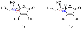
An interesting example is Vitamin C, also known as ascorbic acid. Its formula is C6H8O, but it exists as two enantiomers (mirror-image isomers), "L" (for "Levo" or left) and "D" (for "Dextro" or right). Although the L and the D molecule of vitamin C have exactly the same chemical composition; their properties are drastically different[3], while the L conformation exhibits numerous beneficial healing properties the R isomer is neutral in most cases.


isomer vitamin C ascorbic acid.png

© Yikrazuul
Isomers of vitamin C: L-ascorbic acid (1a); D-ascorbic acid (1b)


Vitamin C is just one example among many others. Two enantiomers of a same compound can significantly differ in their taste, smell or other biological actions.

For example, (+)-limonene found in orange (causing its smell), and (–)-limonene found in lemons (causing its smell), show different smells[4] . The same applies to carvone: (+)-carvone is responsible for the smell of caraway seed oil, whereas (–)-carvone is responsible for smell of spearmint oil.[5]

The same goes for pharmaceutical molecules, two enantiomers sometimes show remarkable difference in terms of biological actions[6]. For instance, D-propoxyphene[7] is a documented painkiller, whereas its enantiomer, L-propoxyphene[8] is an effective anti-cough agent.

In the same vein, the S-isomer of penicillamine, is a treatment of primary chronic arthritis, whereas the R-isomer has no therapeutic effect and is highly toxic.[9]

The preeminence of geometry over composition in defining the properties - informational content - of a substance was explained, until recently, by the key-locks model invented by German chemist Emil Fischer in 1894[10]. According this simple model - chirality matters only on a mechanistic level: the geometric shape of a protein allows or not its binding to a specific receptor. In this context, the “fitting” is mechanical, it occurs between a pattern along the key and the counter pattern within the lock.

But, in today’s technology, remote control keys do not have any pattern fitting at all. The key sends an electronic signal which enables the locking, or the unlocking of the lock. [11]. Could it be same at the cellular scale?

Fortunately, since the 1890’s, then numerous direct and indirect observations of “binding” sites have revealed that the lock and key analogy was a simplistic mechanistic analogy unable to account for numerous documented features of intercellular dynamics:

in terms of vibrations, the human body can be compared to a symphony orchestra. Each molecule corresponds to a particular instrument. Each bend, rotation, or stretch of a chemical bond has a certain resonant frequency and will give off certain frequencies or 'notes' if it is energized. Since molecules, water, and dissolved ions are constantly bumping into each other at body temperature, all parts are constantly jiggling and absorbing and emitting characteristic frequencies.
While a chemical process, such as the breaking of a bond, may look superficially like a mechanical event, at a deeper level the event is better described as a series of vibratory energetic interactions. This is one level at which the various energy therapies may have their effects.
A soprano shatters a crystal goblet by singing a high note coinciding with the natural frequency of the goblet. The atoms in the glass vibrate so strongly that they cannot hold together, and the gobelet breaks. The same thing can happen to a molecule. Figure 9.10 shows a molecule of hydrogen Peroxide, H2O2, being fractured by vibrations. Sometimes this is called 'molecular surgery'.
The oxygen--oxygen (0-0) bond of hydrogen peroxide is broken selectively with an electromagnetic field of a specific frequency. The wavy arrow in the drawing represents a photon of laser light that energizes the O—H bond (i.e., it makes the bond vibrate violently) just as a tuning fork resonates when you strike it. The vibrations are rapidly conducted or redistributed throughout the molecule, and the 0-0 bond, which is weaker than the 0—H bond, breaks.[12]

Random diffusion, which is the biochemical mechanism of action fostered by the lock and key model is simply too imprecise (random) and too slow (diffusion) to account for numerous biological processes:

random diffusion is far too slow and far too imprecise to regulate living processes in an orderly timely manner. Diffusion of regulatory molecules and diffusion of metabolites within cells re processes that are too slow to allow the organism to adapt to rapid changes would take about in its environs- lent' If random diffusion is the mechanism involved in cellular metabolism,1,000 years for you to digest your breakfast! [13]

The lock and key model is a materialist theory based on random mechanical process. In this sense the lock and key model is to biochemistry what Darwinism is to “evolution”.

Random mutation has as little chance to explain evolution - the chance that a tornado sweeping through a junkyard might assemble a Boeing 747, according to Fred Hoyle[14] - as random diffusion can explain biochemistry.

The lock and key model postulates that the random diffusion of a ligand is what directs its binding of the to its receptor. It’s akin to being blindfolded, holding a key, not knowing where the lock is, poking the key here and there, and hoping the key will magically end up in the right position, the right location, in the right lock at once!

At least in some cases, if not in most cases, the biological communication processes don’t follow the imprecise, slow lock and key model but the fast and precise resonance model:


lock and key.jpg

© Oschman
The lock and key model (A) VS the resonance model (B)


The resonance model is not a novelty; it was created by Malcom Dyson in 1937[15]. Since then, this theory has been experimentally confirmed in various fields.

The lock and key model repeatedly failed to predict the potency of a given agonist, on the contrary to the resonance model for which both the shared spectral peak of related agonists and the intensity of this peak are predictors of agonist potency[16]:

Although the lock and key mechanism has been very successful in modelling certain aspects of the action of signaling proteins one of the ways in which it falls short is the prediction of agonist potency. In addition to the shared spectral peak of related agonists Hoehn et al. also report that the intensity of this peak might be used as a predictor of agonist potency[17]

The above suggests that the ability of a protein to interact with a receptor is not its geometric shape but its resonance characteristics.

The resonance model of molecular interactions has been particularly investigated in the domain of olfaction (sense of smell). For example, experiments on fruit flies showed a resonance phenomenon in the neuro-receptors related to smell:

In his 1996 paper Turin suggested, that the vibrational frequency of a given odorant contributes to quantum tunneling at the receptor. In 2011 Franco et al. presented experimental evidence in support of the theory. In particular they claimed that fruit flies could differentiate between deuterated odorants.[18]

A deuterated odorant refers to a molecule where a usual hydrogen atom (one proton + one electron) was replaced by a deuterium atom (one proton + one neutron + one electron).

deuteriumHydrogenIsotope.gif
© NASA
Hydrogen and deuterium atoms

This minute addition of one neutron to the hydrogen nuclei occurs only at the atomic level. It doesn’t change at all the odorant at a molecular level, therefore falsifying the lock and key model, which is based on geometry differences on a molecular level.

The addition of one neutron in the deuterium atom compared to the hydrogen atoms modifies the geometric shape on an atomic level and the whole resonance properties of the molecule[19] constituted by these atoms.

The ability of the fruit flies to smell a difference between deuterated and un-deuterated odorants suggests that the perception of smell is not defined by the composition of the molecule, but the geometric shape at an atomic scale and the subsequent vibrational signature.

In 2001, a study conducted at Purdue University showed that, like fruit flies, humans can also perceive the difference in smell between normal and deuterated versions of a given molecule. [20]

Similar experiments have shown that lake white fish and the American cockroach can differentiate between isotopes of the same amino acids and pheromones[21]. Like in deuterated vs hydrogenated odorants, the difference between isotopes of a same element is only the number of neutrons in the nucleus of the atom.

But, agonist prediction and smell biochemistry are only some of the domains were the resonance model has been successfully tested. Literally thousands of proteins revealed resonance interactions at a distance:

In our previous studies, the above criteria have been tested with over 1000 proteins from 25 functional groups. The following fundamental conclusion was drawn from our studies: one RRM [Resonant Recognition Model] peak frequency characterizes one particular biological function or interaction. Therefore, those peaks are named as the RRM characteristic frequencies. It was shown that proteins and their interacting targets (receptors, binding proteins, and inhibitors) display the same characteristic frequency in their interactions. However, it is obvious that one protein can participate in more than one biological process, i.e. revealing more than one biological function. Despite proteins and their targets have different biological functions they can participate in the same biological process. Therefore, it has been postulated that interacting molecules "communicate" with each other, i.e. recognise each other at a distance, on the basis of the same/similar (within the calculation error) characteristic frequency but opposite phases at that frequency[22]

The cases of resonances in proteins interactions are so prevalent, that after repeatedly analyzing the spectrum of the distribution of the energies of free electrons along proteins backbone, Emiritus professor Irena Cosic concluded:

protein activities (interactions) are based on resonant electromagnetic energy transfer.[23]

From the above we now know that two substances exhibiting the very same composition reveal drastically different properties depending on their geometric shape, whether on a molecular scale as illustrated by chiral molecules or on an atomic scale as exemplified by the isotope experiments.

These properties of a given substance are defined by its specific connection with the information field. In this case, one can state that the properties of a substance are defined by the specific “area” of the information field it is connected to, this specific connection is defined by the geometric shape of the substance, which modulates its resonance signature.

The above shows that while the mechanistic lock and key model can explain some biochemical processes, it fails to explain others for which resonance seems to be involved.

So, in the next chapters, we will attempt to understand how the geometric shape of a given substance is associated with its resonance and with its informational content.

Why two molecules displaying the exact same composition differ so much in properties/informational content? We know that geometric shape obviously plays a fundamental role, but how does it work? Is the difference in geometry altering a fundamental factor involved in modulating the connection between the information field and the proteins? What is this fundamental factor?

The next chapter aims at answering these questions.


[1] Alberts, B. et al. (2002) “Molecular Biology of the Cell. 4th edition”. Garland Science in The Shape and Structure of Proteins
[2] Smith, Yolanda (2018) “Protein Structure and Function” News-Medical
[3] Wu X, et al. (2020) “A chirality-dependent action of vitamin C in suppressing Kirsten rat sarcoma mutant tumor growth by the oxidative combination: Rationale for cancer therapeutics” Int J Cancer 146(10):2822-2828
[4] Solomon and Fryhles. “Organic chemistry”, Ed 10, Wiley (Students edition), Chapter 5 (Stereochemistry), Section 5.5 More about biological significance of chirality
[5] Ibid
[6] Sanganyado, et al. (2017) "Chiral pharmaceuticals: A review on their environmental occurrence and fate processes". Water Research. 124: 527–542.
[7] brand name Darvon
[8] brand name Novrad
[9] Solomon and Fryhles organic chemistry, Ed 10, Wiley (Students edition), Chapter 5 (Stereochemistry), section 5.11(chiral drugs).
[10] Liu, Shu-Qun et al. (2012) “Protein Folding, Binding and Energy Landscape: A Synthesis” in Protein engineering Kaumaya
[11] Arieh Ben-Naim (2018) “Solvent Effects and the Lock and Key Model for Molecular Recognition” Int J Bio & Lab Med. 1:1, 01-06
[12] Eden, Donna & Feinstein, David (1999) “Energy Medicine” Tarcher/Putnam p.137
[13] Ibid, p.133
[14] Gatherer, Derek (2008) "Finite Universe of Discourse: The Systems Biology of Walter Elsasser (1904-1991)" The Open Biology Journal. 1: 9–20
[15] Hoehn RD et al. (2018) “Status of the Vibrational Theory of Olfaction” Front. Phys. 6:25
[16] An agonist is a chemical that “binds” to a receptor and activates the receptor to produce a biological response. In contrast, an antagonist blocks the action of the agonist.
[17] Hoehn RD et al. (2015) “Neuroreceptor activation by vibration-assisted tunneling”. Sci Rep.;5:9990
[18] Adams, B. et al. (2020) “Quantum effects in the brain: A review” AVS Quantum Science. 2. 022901 10.1116/1.5135170
[19] More specifically it adds vibrational modes of the carbon-deuterium bond. See:
A. P. Horsfield et al. (2017). “Molecular recognition in olfaction” Advances in Physics: X, 2:3, 937-977
[20] Haffenden LJ, Yaylayan VA, Fortin J (2001). ‘’Investigation of vibrational theory of olfaction with variously labelled benzaldehydes”. Food Chem. 73 (1): 67–72
[21] Havens BR, Melone CD (1995) “The application of deuterated sex pheromone mimics of the American cockroach, to the study of wright's vibrational theory of olfaction”. Dev. Food. Sci. 37 (1): 497–524
[22] Cosic, I., Pirogova, E. (2007) “Bioactive peptide design using the Resonant Recognition Model”. Nonlinear Biomed Phys 1, 7
[23] Cosic, Irena. (2001) “The Resonant Recognition Model of Bio-molecular Interactions: possibility of electromagnetic resonance” Polish Journal of Medical Physics And Engineering. 7. 73-87
 

Attachments

  • isomer vitamin C ascorbic acid.png
    isomer vitamin C ascorbic acid.png
    18.7 KB · Views: 12

Chapter 5: Biophotons​


We have presented in the previous chapter the concept of chirality. As mentioned previously, molecules with the same composition can present two mirror images: "right-handed" (D) or "left-handed" (L). See the cases of ascorbic acid for example.


Chirality_with_hands.protein.png
© Unknown
The two different chiralities of a molecule

But, what do (D) and (L) exactly stand for? Actually (D) is the initial of Dextrorotatory while (L) is the initial of Levorotatory.

In fact the very definition of dextrorotatory (D) vs levorotatory (L) has nothing to do the chemical properties or chirality per se but with the different ways each of these two types of molecules bent photons:

They're identified by the way they bend light in solution. The dextrorotatory (D) forms rotate it to the right, while levorotatory (L) isomers refract it to the left.[1]

enantiomer-rotate photonsl.jpg
©Libretexts
When polarized light interacts with chiral molecules, the plane of polarization is rotated

More than that, according to Irena Cosic, it seems that photons, both visible and invisible, are directing the very activity of proteins:

Protein activities (interactions) are based on resonant electromagnetic energy transfer within a range of infra-red and visible light.[2]

To illustrate this point let’s take the example of two molecules: 3-4 benzopyrene and 1-2 benzopyrene, which are organic compounds with exactly the same chemical composition C20H12:


benzopyrene.jpg
© Sott.net
Left: 3,4-benzopyrene. Right: 1,2-benzopyrene;


As shown in the diagram above, the only difference between these two molecules is geometrical (location of the benzene compound) but the properties differ drastically. While 3-4 benzopyrene is highly carcinogenic, 1-2 benzopyrene is an harmless molecule.

How can we explain that no difference in chemical composition and only a minute geometrical difference induce such a large difference in chemical properties? When Fritz Popp analyzed the photons emissions of the two molecules he noticed something “unusual”:

The two molecules of 3-4 benzopyrene and 1-2 benzopyrene differ fundamentally above all in the absorption and reemission of ultraviolet light. Thus, 3-4 benzopyrene exhibits an energetic coincidence that must be considered unusual for a molecule, it is a "quasi degeneracy" of the three lowest ultraviolet excitation states.[3]

One reason why this anomaly in the UV light absorption and reemission has such a drastic effect is because the UV band seems to be used predominantly by living cells to communicate with each other:

Three Soviet scientists, S. Stschurin, V.P. Kaznachejev and L. Michailova, confirmed in more than 5,000 experiments that living cells transmit information by photons, and in particular by light in the ultraviolet band. This experiment can be described simply as follows:
Cells in a nutrient solution live in two quartz balloons. The containers touch each other at the wall. One of the cell cultures is contaminated with a virus: almost at the same time, the cells of the adjacent colony also become ill. The same thing happens when the cells are destroyed by doses of ultraviolet light or poisoned by sublimate. The cells in the neighboring container become ill each time and show the same symptoms, although they should be protected from the toxic influence of the other cells by the quartz container. It is only when normal glass is used instead of quartz [ordinary glass absorbs ultraviolet radiation while quartz allows it to pass] that the cells of the second colony are spared the ills suffered by the first. Some disease information from the first colony must therefore pass through the quartz walls and not the glass walls to the second colony. It cannot be the chemicals or viruses introduced into the first culture. These were not found in the neighboring culture...[4]

So this experiment confirms that it is not the material substance (virus or protein for example) that matters per se but the informational content it attaches to. In other words, what matters is not the material substance but what information it connects to in the Information Field.

The above suggests that it’s not the conformation that causes directly the difference in properties. There seems to be one more step: the geometry affects the photons that in turn affect in the chemical properties (informational content) of a given molecule:


geometry biophotons with information field.jpg
© Sott.net
Geometry modifies light properties that modifies the informational content
that in turn modifies chemical properties

We hypothesized above that the light properties affect our connection with the information field. If this is true, one question is how photons can carry information and, at the same time, be compatible with proteins “antennas”, knowing that these protein antennas display fractal properties:

Fractal nature of protein surface roughness: a note on quantification of change of surface roughness in active sites, before and after binding[5]

An individual photon is defined by a few parameters like its wavelength, amplitude and diffraction mode, so its information-carrying capacity is limited proportionally its number of parameters. In addition, an individual photon doesn’t display fractal patterns that would match with the protein/fractal antenna.

Logically as a group, photons can communicate much more information than an individual photon. But this information carrying capacity is de-multiplied if the group of photons behaves in a coherent manner like a Laser for example. In this case synergies appear and the group of coherent photons can carry way more information than the sum of its parts (individual photons). Here is an example that illustrates the above point:

An infrared laser can carry information in a similar way to radio waves. By modifying the invisible beam a certain way, the varying modulation can transmit a digital signal […] In 2013, a demonstration on the Lunar Atmosphere and Dust Environment Explorer (LADEE) spacecraft relayed video between groundstations on Earth and the orbiter. The orbiter was able to transmit 622 megabits of data, enough to carry 30 HDTV channels[6]

But do biophotons display coherence like Lasers do? Several prominent researchers in the field of biophotons claim so, among whom Bischof[7], Popp and Yan[8], and Bajpai[9] [10].

As stated in the quote above, coherent photons display amazing information carrying capacity but what about the match between the signal (photons) and the antenna (proteins)? We know that proteins contain fractal patterns; can coherent photons also produce fractal patterns?

Light produced by Lasers is coherent; it means in layman’s terms that photons move in unison or in more technical terms the photons wavelengths are in phase in space and time as described in the illustration below:
coherent waves laser .jpg
© Unknown
Photons produced by a laser are coherent

In 2019, for the first time, scientists managed to produce fractal light patterns from lasers, i.e. coherent photons[11]. In addition they've shown the fractal light could be created in 3D rather than just in 2D:


laser fractals.jpg
© Wits University
Fractal light created by Laser

Like lasers, structured water in living creatures is coherent:

The reason living organisms could appear like a dynamic liquid crystal display is because all of the molecules in the tissues and cells including especially the water molecules, are not only globally aligned as liquid crystals, but also moving coherently together as a whole.

Here is a short video made Mae Wang Ho[12]. It shows how water in a live being is a liquid crystal; it diffracts light like a solid crystal prism. Notice that when the lens of the microscope is rotated the colors of the same body part of daphnia change:


Furthermore structured water is known to emit biophotons[13]. So, one of the next logical questions is: Do biophotons emissions reveal fractal pattern?

As a matter of fact, they do. Although the topic of biophotons in conjunction with fractals is seldom investigated, fractal patterns have been shown in biophotons emissions in this paper titled Short-Time Fractal Analysis Of Biological Autoluminescence:

Results from rigorous statistical analysis and testing in Tables 3,4, and 5 suggest that the mung beans signal has a negative memory (negative correlations, antipersistent behavior) and its Hurst exponent is lower than the referential signal. How could such behavior originate in biological systems? It was proposed that the restriction of Brownian motion due to the structuring of nano- to microscale intracellular environment leads to anomalous sub-diffusion characterized by Hurst exponent < 0.5. This is understandable since a cytoplasm environment displays fractal spatial structuring. Since biochemical reactions (encounters of reactants) leading to BAL are taking place within the cell cytoplasm, organelles and lipid membranes [24] where anomalous sub-diffusion was observed [64, 66], it is not a great logical leap to speculate that BAL from mung bean samples could also display sub-diffusive features. Actually, it is already acknowledged that chemical reactions spatially constrained on the microscopic level may lead to fractal reaction kinetics […]
The resulting Hurst exponent[14] mean value of mung bean sample time series is below the level of 1/2 which confirmed our initial hypothesis[15]

To summarize, we saw that two chiral molecules, two proteins for example, have exactly the same composition; they only differ in handedness. The difference in chirality leads to different biophotons emissions enabling connections the different area of the Information Field, that in turn confer the two molecules with two different informational content i.e. chemical properties:


biophoton emission protein fractal.jpg

© Sott.net


The above suggests how important epigenetic factors (diet, emotions, thoughts, actions...) are; for these changes, DNA will express this or that protein/antenna. This change in proteins induces a change in biophotons emissions (fractal patterns) that modulates your very connection to the information field; literally modifying the information you access to.

Here is a diagram showing how chromatin can activate or de-activate gene expression (production of this or that protein/antenna):


Open-and-closed-chromatin-configurations-are-influenced-by-post-translational-histone.jpg

© Gibney
Chromatin can suppress the expression of a gene by neutralizing histones

Open and closed chromatin configurations are influenced by post-translational histone modifications. In the upper panel, DNA is wrapped around histones that possess activating modifications (green circles and blue triangles). In the lower panel, DNA is wrapped around histones with repressing modifications (red circles and orange triangles). The bent arrow indicates a transcription start site; this is more accessible to RNA polymerase in the open chromatin configuration.[16]

Coincidently or not, notice that the same chromatin which plays a crucial role in gene expression has also chromatin has photoreception properties:

Cell Type-Specific Epigenomic Analysis Reveals a Uniquely Closed Chromatin Architecture in Mouse Rod Photoreceptors[17]

At this point, you might wonder where biophotons ultimately come from, how are they produced? It seems that oxygen is the main source of biophotons:

The emergence of biophotons is due to the bioluminescent radical and nonradical reactions of Reactive Oxygen Species (ROS)[18]
More precisely it is the change in electrons spins around the molecule of dioxygen that creates biophotons:

Following a complicated reaction scheme that requires a second oxygen molecule, the spin-saturated complex breaks down again into two spin-coupled O2 molecules in parallel with light emission. 2 OS2 → 2 OT2 + h υ. The transition from oxygen dimolecular singlet to triplet thus produces photons with a wavelength of 634.7 nanometers (1 nm = 10-9 m)[19]

oxygen spin triplet singlet.jpg
© Unknown
The transition from O2 singlet to triplet produces photons

In conclusion, we can hypothesize that epigenetic changes may modulate our connection to the Information Field while oxygen may strengthen it.


[1] Robert Becker, Gary Selden (1998) “The Body Electric: Electromagnetism And The Foundation Of Life” William Morrow Paperbacks
[2] Cosic, Irena (2001) “The Resonant Recognition Model of Bio-molecular Interactions: possibility of electromagnetic resonance” Polish Journal of Medical Physics And Engineering 7. 73-87
[3] Fritz-A.Popp (1984) ‘’Biologie de la Lumière’’ Résurgence
[4] Popp, 1984
[5] Banerji A, Navare C. (2013) “Fractal nature of protein surface roughness: a note on quantification of change of surface roughness in active sites, before and after binding” J Mol Recognit. 26(5):201-14
[6] Andrew Wagner (2020) ‘’Communicating via Long-Distance Lasers” NASA
[7] Bischof, Marco (2005) “Biophotons- The Light in Our Cells” Journal of Optometric Phototherapy 15. 1-5
[8] Popp F.A. & Yan Y. (2002) “Delayed luminescence of biological systems in terms of coherent states” Physics Letters A. 293. 93-97
[9] Bajpai, RP. (1998). “Coherent Nature of Biophotons: Experimental Evidence and Phenomenological Model” in: Biophotons. Springer
[10] Bajpai RP. (1999) “Coherent nature of the radiation emitted in delayed luminescence of leaves” J Theor Biol. 198(3):287-99
[11] Hend Sroor et al. (2019) “Fractal light from lasers” Phys. Rev. A 99, 013848
[12] Mae-Wan Ho (2008) “The Rainbow and the Worm: The Physics of Organisms” World Scientific
[13] Glen Caulkins (2012) ‘’ Biophoton Energy Water’’ Personal website
[14] A Hurst exponent below 0.5 means a fractal dimension comprised between 1.5 and 2
[15] Dlask M. et al. (2019) “Short-time fractal analysis of biological autoluminescence” PLoS ONE 14(7): e0214427
[16] Gibney, E. & Nolan, C.M. (2010) “Epigenetics and gene expression” Heredity 105. 4-13. 10.1038
[17] Hughes, A. et al. (2017) “Cell Type-Specific Epigenomic Analysis Reveals a Uniquely Closed Chromatin Architecture in Mouse Rod Photoreceptors” Sci Rep 7, 43184 (2017)
[18] Rahnama, Majid et al. (2010) “Emission of Biophotons and Neural Activity of the Brain” Arxiv
[19] Popp, 1986
 
C'est formidable tout ce que vous faites, découvrez, expliquez mais mon intelligence n'est pas suffisante pour vous suivre...
Je vous remercie car tout le mal que vous vous donnez pour nous servira à d'autres, j'en suis certaine...

It's great what you do, discover, explain but my intelligence is not enough to follow you...
I thank you because all the trouble you go through for us will be useful to others, I am sure...
 
Here is the pdf for chapter 5.
For anyone else who’s interested in reading about the triplet state of oxygen and how it effects which biophotons are released there’s information in the book Bioenergetics by Albert Szent Gyorgyi. He was another Nobel Prize winner who was ultimately silenced by the scientific community.

 
Hello Pierre,
I have only read chapter 4 so far.
If I understand correctly, this is the scientific demonstration of various exchanges that you have had with the Cs concerning proteins as antennas of the information field? Can we also say that proteins allow an increase in our vibrational frequency if we consider that: "Everything below is like everything above. (Emerald table)?
 
Hello Pierre,
I have only read chapter 4 so far.
If I understand correctly, this is the scientific demonstration of various exchanges that you have had with the Cs concerning proteins as antennas of the information field?

Correct

Can we also say that proteins allow an increase in our vibrational frequency if we consider that: "Everything below is like everything above. (Emerald table)?

The proper proteins might enable an increase in FRV, but that is just speculations on my part.
 
Back
Top Bottom