Basic Anatomy
The nervous system consists of two main divisions: central and peripheral.
The central nervous system (CNS) includes the cerebrum (A.K.A brain or cortex), cerebellum, brain stem, and spinal chord plus a few scary-sounding structures located between the brain stem and the brain, the diencephalon (which includes everything with the name “thalamus”; i.e. the thalamus, hypothalamus) and the basal ganglia (which includes the amygdala among others).
The basic functional unit in the CNS is the neuron. The electrical impulse travels down a neuron from its dendrites to the cell body and axon. Information then is chemically transmitted to other neurons via connections known as synapses. A chain of such communicating neurons is called pathway. Within the CNS, a bundle of pathway axons is called a tract, fasciculus, etc. Outside the CNS, bundles of axons are called nerves. As you can see, there are many different names to say the same thing.
Axons conduct the electrical impulse, the speed of conduction depending on fiber diameter. But efficiency of conduction also depends on good insulation, which is achieved by the myelin sheath. The myelin sheath coats the axon’s membrane and it is composed mainly of fat.
The brain is subdivided into frontal, parietal, occipital and temporal lobes. These are further subdivided into bulges, called gyri, and indentations called sulci and fissures.
The brain stem contains three parts – the mid brain, pons, and medulla. The word “bulbar” refers to the brain stem and all its three parts. Bulbar comes from bulb and if you look at it, it does resemble a bulb from a plant. Keep that in mind when you hear about corticobulbar pathway.
Brain anatomy is very logical. You just have to look at a word to see what is telling you. Cortico comes from cortex (brain). Bulbar refers to the brain stem. So it is the pathway that goes from the brain to the brain stem.
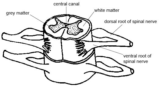
The spinal chord contains central grey matter and peripheral white matter. The grey matter is rich in neurons. The white matter contains ascending and descending pathways corresponding to sensory and motor pathways respectively. The ascending pathway relays incoming information (sensory information- from senses, i.e. oooh, ice is really cold!) to the brain. All sensory pathways end up in the thalamus. From there, they are relayed to the sensory area of the brain.
The descending motor pathway (which allows us to kick something) is called the corticospinal pathway. Cortico comes from cortex, the brain. Spinal is the spinal chord. That means that it is the pathway that goes from your brain to your spinal chord, which then will relay information to the nerves in your leg that will make you kick something if you want to.
The corticospinal pathways synapses in the motor grey matter (which is called the “anterior horn”) of the spinal cord just prior to leaving the cord. Motor neurons above the level of this synapse (connecting the brain and anterior horn) are termed upper motor neurons, whereas those beyond this level (the peripheral nerve neurons) are termed lower motor neurons. It might sound complex, but it just a very specific way to say where is “up” and where is “down”.
As tedious as it might be, it is necessary because it is so specific that it is almost mathematical. When you are talking about an upper neuron or a corticobulbar pathway, it refers to something specific and nothing else. There is no space for misunderstanding or misinterpretation.
These motor and sensory pathways belong to the somatic motor (innervating skeletal muscles) and somatic sensory (innervating skin, muscle and tissues other than the viscera) systems. Somatic is just a fancy way to say body-related.
Within the context of the nervous system, afferent refers to sensory fibers that carry information to the brain, and efferents carry information away from it (i.e. motor efferent fibers that carry information from the brain to a muscle or organ).
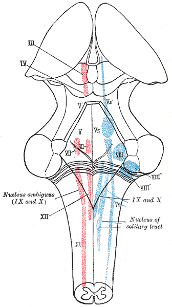
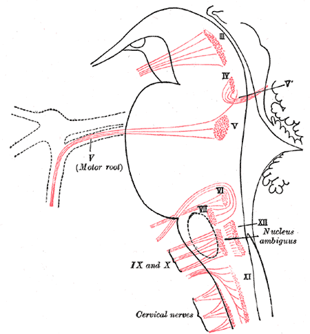
The brain stem contains anatomic groupings of cell bodies known as nuclei which belong to cranial nerves, that is, nuclei is where cranial nerves like the vagus nerve will arrive to (afferents of the cranial nerves) or will originate from (efferents of the cranial nerves). Nuclei serve also as relay cells of ascending sensory or descending motor systems. The remaining cell groups of the brain stem, located in the central part (the grey matter), constitute a diffuse-appearing system of neurons with widely branching axons, known as the reticular formation.
The reticular formation creates a network called the reticular activating system which maintains attention and alertness. A difficulty in the control of attention may contribute to major symptoms associated with psychopathy. [A.R.Baskin-Sommers, J.D. Zeier, J.P. Newman. Self-reported attentional control differentiates the major factors of psychopathy. Personality and Individual Differences 47 (2009) 626–630.]
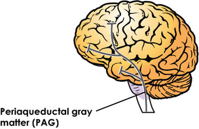
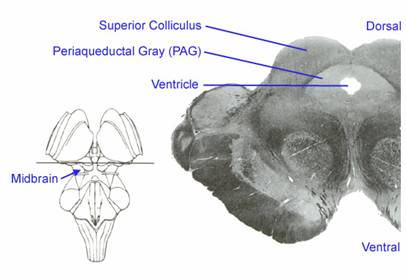
Opiate-like activity chemicals (endorphins) and their receptors have been found in various areas of the reticular formation including the grey matter surrounding the aqueduct, a canal running through the brain stem (hence its name, periaqueductal grey matter).
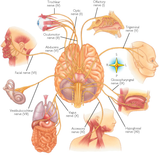
The peripheral nervous system (PNS) consists of cranial and spinal nerves. There are 12 pairs of cranial nerves from which the vagus nerve is the 10th.
The vagus is the heart of the parasympathetic system and it has extensive motor and sensory components with nerve trunks originating in the left and right sides of the brainstem. The vagus nerve is asymmetrical, with the left and right sides performing different tasks, with the right vagus having the most important role in heart rate determination. Efferent motor fibers from the vagus nerve originate primarily in two nuclei at the medulla (in the brain stem) called nucleus ambiguus and dorsal motor nucleus of the vagus. Afferent sensory fibers from the vagus nerve (80% of all the fibers of the vagus nerve) end up in the nucleus tractus solitarius in the medulla (brain stem).
The Autonomic Nervous System
The Autonomic Nervous System (ANS), is the autonomic portion of the nervous system that takes place without conscious action on our part. It has to do with all those functions in our bodies that are involuntary, “unconscious”, i.e. the viscera. “Viscera” refers to heart muscle, smooth muscle (as in the gut) and glands.
The word autonomic comes from the root auto (meaning “self”) and nomos (meaning “law”).
Thanks to the ANS, our bodies respond automatically to changes in the physical or emotional environment. For example, when our bodies respond automatically with freaky sensations in response to a horror movie we are under the control of the ANS. Indeed, the autonomic nervous system is a marker of emotional activity.
The ANS is usually divided in two: sympathetic and parasympathetic. These two divisions have brain chemical differences and they also differ in embryological origin (from embryo or fetus. That is, a baby being formed in his mother’s womb) and function.
In the old theory, the one that is still quoted in textbooks and pretty much everywhere, specific physiological reactions (including brain reactions) following stress are often associated with the sympathetic nervous system and the hypothalamic-pituitary-adrenal (HPA) axis. [In the polyvagal theory we’ll learn] that this is not necessarily the case.
The sympathetic system as a whole is generally associated with a catabolic system, that is, expending energy, as in flight or fight response to danger. The parasympathetic system is an anabolic system, conserving energy. Parasympathetic synapses typically lie very close to or within the viscera. The final synapse of the parasympathetic’s system contains the neurotransmitter acetylcholine, whereas the final synapse of the sympathetic system contains noradrenaline (a stress hormone), with the exception of certain synapses, as for sweating, that contain acetylcholine (i.e. are cholinergic).




 And the price doesn't matter just a difference of 2-3 Euros to the original English (hardcover) book, the German (about 35€) one is a softcover.